Ankle Sprains: Don’t Let a Simple Injury Turn into a Long-Term Problem

Ankle Sprains: Don’t Let a Simple Injury Turn into a Long-Term Problem

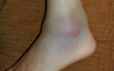
Ankle sprains are among the most common injuries we see at Praxis Physiotherapy. Whether you’re an AFL midfielder, a cricket fast bowler, or a weekend runner pounding the Brisbane River loop, lateral ankle sprains can derail performance and linger longer than they should.

At Praxis, we’ve rehabilitated hundreds of athletes across all levels, from juniors to pros. Our experience includes long-term roles with the Aspley Hornets AFL Club (since 2014), the Queensland Bulls, Australia A, and even the Australian Men’s Cricket Team. We bring these elite rehab principles to everyone — from sprained-ankle soccer kids to high-performance track athletes.

But despite how common they are, ankle sprains are often underestimated. Without proper rehab, they can lead to chronic ankle instability (CAI), impaired athletic performance, and even new injuries in other parts of the body.

What Actually Happens in an Ankle Sprain?

A lateral ankle sprain usually occurs when the foot rolls inward, stretching or tearing the ligaments on the outside of the ankle — most commonly the anterior talofibular ligament (ATFL). It often happens during sudden changes of direction, awkward landings, or stepping on uneven ground.

You might feel a pop or crunch, followed by swelling, bruising, and pain when walking or bearing weight. While it may seem like a “simple sprain,” it’s anything but — around 40% of people report long-term issues one year post-injury if not managed well​.

gray concrete statue of a man

Common Mistake: Rest, Ice, and… That’s It?

Too many people still follow the old R.I.C.E. (rest, ice, compression, elevation) model and assume the job is done. While these strategies can help in the first 48 hours, they’re far from sufficient for full recovery.

In fact, research has shown that inadequate rehab is a major contributor to chronic ankle instability — a condition marked by recurrent sprains, feelings of the ankle “giving way,” and reduced confidence in movement​.

CAI can lead to altered biomechanics and poor neuromuscular control, increasing the risk of knee injuries, Achilles tendinopathy, or even hip and low back pain due to compensation.

Proper Rehabilitation Is Key — Here’s What the Evidence Says

Rehabilitation needs to start early and be progressive. High-quality clinical guidelines and systematic reviews strongly support the following strategies:

Functional Support and Early Mobilisation

Functional bracing (like an ankle brace or taping) is preferred over rigid immobilisation and should be used for 4–6 weeks . Early weight-bearing as tolerated leads to quicker return to activity and better outcomes .

Exercise Therapy

Neuromuscular training (balance, proprioception, and strength work) is the foundation of successful rehab. It improves ankle control, prevents recurrence, and reduces the risk of CAI​. A wobble board, single-leg balance, hopping drills, and directional change exercises are all commonly used.

Manual Therapy

Joint mobilisations and soft tissue work may improve dorsiflexion range, decrease pain, and aid in functional recovery​. At Praxis, we combine manual therapy with functional retraining to fast-track performance readiness.

Individualised Return-to-Sport Testing

Return to sport shouldn’t be based on time alone. We use objective testing — including single-leg hop symmetry, balance tests, and strength assessments — to ensure you’re not returning with deficits that could increase your reinjury risk.

The Cost of Incomplete Rehab: What Happens If You Don’t Get It Right?

A rushed or poorly structured rehab may get you back to activity temporarily — but it opens the door to:

  • Chronic Ankle Instability (CAI): Repeated sprains, perceived instability, and loss of ankle confidence.

  • Performance Limitations: Reduced agility, speed, and power due to poor proprioception and strength deficits.

  • New Injuries: Compensatory patterns can lead to medial tibial stress syndrome (shin splints), Achilles overload, or even ACL risk due to poor landing mechanics.

In elite sport, we see this cascade far too often. That’s why our rehab at Praxis isn’t just about the ankle — it’s about restoring whole-limb function and confidence under pressure.

Prevention: Keep Your Ankles Bulletproof

At Praxis Physiotherapy, we don’t just treat ankle sprains — we help prevent them. Our prevention approach includes:

  • Regular Balance and Plyometric Training: Incorporating single-leg exercises into gym and field work.

  • Proprioceptive Work: Using wobble boards, balance mats, and directional hopping.

  • Footwear and Bracing Advice: Particularly for high-risk sports like netball, football, and athletics.

  • Pre-season Screening and Performance Testing: For our affiliated sports clubs and athletic populations.

Evidence supports proprioceptive training as a proven strategy to reduce ankle sprain incidence by up to 35% in high-risk athletes​.

Why Choose Praxis Physiotherapy?

Our exposure to elite sport has taught us what good rehab looks like — and we apply those same high standards to every patient. Our clinics are equipped with strength testing tools, reformer Pilates, and full gym access, giving you the tools to rebuild better.

We understand the mindset of athletes — from juniors chasing state squads to elite-level players returning from surgery. That’s why we tailor your program based on sport demands, movement patterns, and individual goals.

Whether you rolled your ankle playing touch footy or twisted it at work, we’re here to get you back — stronger, faster, and more confident than before.

Need Help with an Ankle Sprain?

If you’ve recently rolled your ankle or are dealing with ongoing instability, book a consultation at Praxis Physiotherapy. Let our team guide you through a structured rehab program grounded in sports science and elite clinical standards.

📍 Clinics in Teneriffe, Buranda, and Carseldine
💪 Trusted by athletes. Backed by evidence. Here for every body.

References

Ruiz-Sánchez et al. (2022). Management and treatment of ankle sprain according to clinical practice guidelines: A PRISMA systematic review. Medicine (Baltimore), 101(42)

Green et al. (2019). What is the quality of clinical practice guidelines for the treatment of acute lateral ankle ligament sprains in adults? BMC Musculoskeletal Disorders, 20(394)

Doherty et al. (2017). Treatment and prevention of acute and recurrent ankle sprain: an overview of systematic reviews with meta-analysis. BJSM, 51(2), 113–125.

Achilles Tendinopathy: How to treat your Achilles Pain

Achilles Tendinopathy: How to treat your Achilles Pain

Today on the Praxis What We Preach blog, where we shed light on Achilles tendinopathy, a common condition affecting athletes and active individuals. In this article, we will explore the causes, symptoms, and effective treatment strategies for managing Achilles tendinopathy, empowering suffers to return to the things. I draw from personal experience from someone who has had Achilles pain limit my running!

Achilles tendinopathy refers to the degeneration or overload of the Achilles tendon, the band of tissue connecting the calf muscles to the heel bone (calcaneus). This condition primarily affects people engaged in activities involving repetitive jumping, running, or sudden increases in training intensity. Patients with Achilles tendinopathy often experience pain, stiffness, and swelling in the achilles, which can gradually worsen over time. Stiffness and pain is most commonly experienced first thing in the morning, after a long period of sitting or when the achilles has been compressed. Pain can occur in the “mid portion” (pictured below) on in the insertion (as it attaches to the heel bone). This is in an important distinction as these are rehabilitated differently!

Mid Potion Achilles Tendinopathy Location

Causes and Risks

Achilles tendinopathy typically results from a combination of intrinsic and extrinsic factors. Intrinsic factors include age, reduced flexibility, reduced calf strength / endurance and poor lower limb biomechanics. Extrinsic factors encompass inappropriate footwear, training errors (such as a spike or change in workload), and inadequate warm-up or cool-down routines. Additionally, individuals with systemic conditions like diabetes or rheumatoid arthritis may be more prone to developing Achilles tendinopathy. Understanding these factors is crucial for tailoring treatment plans to address the root causes and minimize the risk of recurrence. But in the most reductionist of terms, Achilles tendinopathy develops due in large part due to a mismatch between loading and the capacity of the tissue.

Diagnosis and Assessment

Accurate diagnosis of Achilles tendinopathy relies on a thorough clinical examination and patient history. Physiotherapists employ various assessment techniques, such as palpation, functional tests, and imaging modalities like ultrasound or MRI, to evaluate the severity and extent of the condition. A self administered questionnaire (VISA-A) can help evaluate symptoms and their effect on physical activity and in turn, the clinical severity. This comprehensive assessment helps determine the appropriate treatment approach, including targeted exercise programs, manual therapy, and other interventions.

Treatment Strategies

Physiotherapy plays a pivotal role in the management of Achilles tendinopathy. Treatment strategies focus on reducing pain, promoting healing, and improving function. These will include calf strengthening exercises, stretching routines and activity modification as frontline options. Moreover, physiotherapists can guide patients in proper footwear selection, gait retraining, and implementing preventive measures to minimize the risk of reinjury.

Rehabilitation and Prevention

Rehabilitation programs are essential for individuals recovering from Achilles tendinopathy. Gradual progression of exercise intensity, functional training, and sport-specific drills enable patients to regain strength, flexibility, and proprioception while minimizing the risk of relapse. Educating patients on proper warm-up and cool-down routines, appropriate footwear selection, and regular monitoring of training loads can significantly contribute to preventing Achilles tendinopathy in the future. One of the common errors patients make is making rehabilitation too easy, or returning to sport too quickly. Again, physiotherapy play a pivotal role in ensuring you undertake a graduated return to loading as the application of mechanical stress to the Achilles tendon promotes tendon healing and remodeling.

Conclusion

Achilles tendinopathy requires a comprehensive approach for effective management. As physiotherapists, our knowledge and expertise are invaluable in helping you overcome this condition and return to their active lifestyles. To discuss your Achilles issues with us to get you back to what you love doing, book online with Praxis today.

Until next time, Praxis What Your Preach.

Team Praxis

Plantar Fasciopathy: Understanding how to heal your heel pain

Plantar Fasciopathy: Understanding how to heal your heel pain

Feel like your walking on glass in the mornings?  Those first few steps after a long period of sitting hurt the underside of your heel? Struggling to stand at the end of a long day due to your feet? If so, then you may have plantar fasciopathy, also known as plantar fasciitis. Plantar fasciopathy is a common condition that affects the plantar fascia – a thick band of connective tissue on the bottom of the foot. Plantar fasciopathy commonly affects individuals between the ages of 40 and 60, but can affect almost anyone. In this article, we will delve into the causes, symptoms, treatment options, and preventive measures to help you understand, and more importantly manage, this condition.

Causes and Symptoms

Plantar fasciopathy is often caused by repetitive strain or excessive loading of the plantar fascia, leading to microtears and inflammation. Factors such as overuse, improper footwear, high-impact activities, flat or high-arched feet, and tight calf muscles can contribute to its development. The condition is characterised by sharp pain or a dull ache on the underside of the heel or along the arch of the foot. Pain is typically worse in the morning or after periods of inactivity, and may improve with movement. Standing for long periods or walking on hard floor can also be aggravating.

Treatment Options

The treatment of plantar fasciopathy focuses on reducing pain, promoting load tolerance, and addressing the underlying causes. Physiotherapy interventions play a crucial role in managing this condition. Therapeutic techniques such as manual therapy, stretching exercises, and strengthening exercises can help relieve pain, improve flexibility, and restore foot function. Specifically, improving the windlass mechanism (a phenomena that refers to the tightening of the plantar fascia during the push-off phase of walking or running when you big toe extends). This mechanism helps distribute forces evenly throughout the foot and reduces strain on the plantar fascia. More generally, improvement of the footy intrinsics and plantar flexors more generally have been shown to reduce the severity and duration of symptoms as well.

Additionally, the use of orthotics, taping, or night splints may provide support and alleviate symptoms. Extracorporeal shockwave therapy (ESWT) and ultrasound therapy are also viable treatment options in some cases. In severe or persistent cases, corticosteroid injections or surgery may be considered, though this is usually reserved for when conservative measures have failed.

Preventive Measures

Prevention is key to reducing the risk of plantar fasciopathy starting in the first instance. If you are keen to ‘pound the pavement’ for example, then gradually increase activity levels. Avoid sudden changes in intensity or duration to prevent overloading the foot. This may mean dancing long bouts for the first time in a while, or returning to running post injury. Wear footwear that provides adequate arch support and cushioning. Understand the importance of regular stretching exercises for the calf muscles and plantar fascia.

As physiotherapy professionals, we understand that addressing the symptoms of plantar fasciopathy early is essential for providing effective care. At Praxis, effective care means arming you with adequate advice and education so you can help manage the symptoms yourself. Further, implementing appropriate treatment options and emphasizing preventive measures, we support individuals in overcoming foot pain and restoring quality of life. After all, we aim to Prevent, Prepare, Perform! So if you have heel pain that is stopping you from doing what you would like to do, discuss it with our knowledgeable team today!

Until next time – Praxis What You Preach!

📍 Clinics in Teneriffe, Buranda, and Carseldine

💪 Trusted by athletes. Backed by evidence. Here for everyone.

Understanding Sever’s Disease: A Common Foot Condition in Active Growing Children

Understanding Sever’s Disease: A Common Foot Condition in Active Growing Children

Understanding Sever’s Disease in Growing Children

Sever’s disease — medically known as calcaneal apophysitis — is a common heel condition affecting active children, especially during growth spurts. Despite its name, it’s not a “disease” in the traditional sense, but an overuse injury of the growth plate at the back of the heel.

What Causes Sever’s Disease?

Sever’s disease develops when repetitive stress irritates the growth plate in the heel bone (the calcaneal apophysis). During adolescence, especially between ages 8–15, the heel bone may grow faster than the surrounding muscles and tendons, causing excessive tension at the Achilles insertion site. When coupled with repetitive impact — such as running, jumping, or playing on hard surfaces — this mechanical overload leads to inflammation and pain.

It’s especially common in sports like soccer, basketball, AFL, netball, and gymnastics. Kids going through growth spurts, or who are highly active without sufficient recovery, are most at risk. Tight calf muscles, poor footwear, and biomechanical factors like flat feet or poor shock absorption may also contribute.

Common Symptoms

The main symptom is heel pain that worsens during physical activity and settles with rest. Children may complain of:

  • Pain or tenderness at the back or underside of the heel

  • Limping or toe-walking, particularly after sport

  • Discomfort when pressing on the heel or squeezing it from both sides

  • Stiffness first thing in the morning or after periods of inactivity

Symptoms are usually one-sided but can be bilateral. If left unaddressed, the pain can start to interfere with participation in sport and physical education at school.

Diagnosis

A diagnosis is usually made through clinical history and physical examination by a physiotherapist. Key indicators include heel pain during activity, recent growth, and tenderness at the posterior heel. The “squeeze test” — applying gentle pressure to both sides of the heel — is often positive.

Imaging (X-ray or MRI) is rarely needed unless symptoms persist longer than expected, or there is suspicion of another diagnosis. Importantly, a visible growth plate on X-ray in this age group is normal and not a reason for concern in itself.

Treatment and Management

Treatment is focused on reducing inflammation, offloading the heel, and supporting the child’s return to normal function. It is important to reassure both child and parent that this is a temporary, self-limiting condition.

Key management strategies include:

  • Load modification: Avoiding or reducing high-impact activity is key, especially sports with frequent jumping or sprinting. Your physio can help create a weekly plan to reduce flare-ups while keeping your child engaged and active.

  • Ice: Icing the heel after sport can reduce inflammation and pain, especially in the early stages.

  • Heel lifts or orthotics: Studies, including the 2016 randomised trial by James et al., show that both orthotic devices and cushioned heel lifts can effectively reduce heel stress. These inserts help absorb shock and reduce Achilles tendon tension.

  • Calf stretching and strengthening: Tight calf muscles increase load on the heel. Scharfbillig et al. (2008) emphasised the role of flexibility programs, particularly eccentric calf training, in improving outcomes.

  • Footwear advice: Supportive, well-fitted athletic shoes are essential. Avoid barefoot running or flat-soled footwear during recovery.

  • Manual therapy and taping: In some cases, hands-on techniques and taping methods may be used to reduce load on the Achilles insertion.

According to the trial by Weert et al. (2016), physical therapy combining load management and exercise-based rehab was just as effective as orthotic devices. This supports a flexible treatment approach tailored to the child’s specific needs and activity level.

Medication: Short courses of anti-inflammatory medication such as ibuprofen can help in more severe cases, especially when pain interferes with sleep or daily function. However, these should always be used under medical advice.

Prevention and Long-Term Outlook

The prognosis for Sever’s disease is excellent. Most children recover fully once the growth plate fuses — typically by age 15 for boys and 13 for girls. The condition does not cause permanent damage, though symptom duration can vary from a few weeks to several months depending on activity levels and adherence to management.

To reduce the risk of recurrence:

  • Encourage a proper warm-up and cool-down routine

  • Ensure sport participation is balanced with adequate rest

  • Maintain calf flexibility and foot strength

  • Use shock-absorbing shoes or orthotics during growth spurts

  • Avoid large increases in training volume or intensity

It’s also helpful to educate kids and parents that some discomfort during growth phases is normal, but persistent pain warrants a review. At Praxis Physiotherapy, our clinicians are experienced in managing growing athletes — and ensuring they don’t miss more game time than necessary.

Summary

Sever’s disease is a common and manageable cause of heel pain in growing children. Early recognition, temporary load reduction, and a guided rehab program can ensure a smooth recovery and quick return to sport. If your child is experiencing heel pain that isn’t improving with rest, book in with the friendly and knowlegable team at Praxis Physiotherapy for a tailored management plan.

Until next time, Praxis What You Preach

📍 Clinics in Teneriffe, Buranda, and Carseldine

💪 Trusted by athletes. Backed by evidence. Here for everyone.


References

James, A. M., Williams, C. M., & Haines, T. P. (2016). Effectiveness of footwear and foot orthoses for calcaneal apophysitis: a 12-month factorial randomised trial. British Journal of Sports Medicine, 50(20), 1268–1275. https://doi.org/10.1136/bjsports-2015-094986

Scharfbillig, R. W., Jones, S., & Scutter, S. D. (2008). Sever’s Disease: What Does the Literature Really Tell Us? Journal of the American Podiatric Medical Association, 98(3), 212–223. https://doi.org/10.7547/0980212

Weert, H. C., van Dijk, C. N., & Struijs, P. A. (2016). Treatment of Calcaneal Apophysitis: Wait and See Versus Orthotic Device Versus Physical Therapy. Journal of Pediatric Orthopaedics, 36(2), 152–157. https://doi.org/10.1097/BPO.0000000000000417

Tibialis Anterior – The missing link to pain free legs and performance?

Tibialis Anterior – The missing link to pain free legs and performance?

Shin splints? Painful knee with jumping? Recurrent ankle sprains? These are the types of injuries we fix day in and day out at Praxis Physio. There are number of recommended paths for rehabilitation in theses injuries backed by the research and our clinical experience, but has the evidence been missing something? There is some social media traction in the fitness and exercise world recently around tibialis anterior (TA) loading as a ‘cure all’ for every lower limb injury. Being the physio nerds that we are, we looked in to it for you!

The tibialis anterior is located on the front (anterior) portion of the shin (tibia) – hence the name. Its role is to lift the foot (dorsiflex) the ankle as well as provide some stability for the outside of the ankle . This action is key for movements in walking and running especially in negotiating steps and hills as the foot needs to clear a certain height before landing. It is also very important in landing from a height and changing directions rapidly, as it acts as shock absorber for the knee and ankle joints (reference).

Like any muscle or joint in the body, the TA is not without its problems. Physios are regularly confronted with patients who complain of pain and stiffness around the muscles of the shin. Such conditions may include shin splints, compartment syndrome, patellofemoral pain syndrome (aka runner’s knee) and general ankle joint pain and stiffness post ankle sprain.

As mentioned, there has been a lot of discussion lately in the strength and conditioning community about whether training this muscle can prevent or treat musculoskeletal conditions, such as the ones mentioned above. Anecdotally, training of the TA has been described several benefits. Specific athletes have explained that they have seen improvements in vertical jump height, running speed, running stamina and squat depth. From a prevention and treatment point of view, it has been said that training the TA has helped improve symptoms associated with knee pain, ankle pain and shin splints. Also let us not forget, that from an aesthetics point of view, a strong and bulky looking TA does make our legs look more attractive, as bodybuilders would argue.

Unfortunately, the scientific community has not provided strong evidence that training the TA can aid in affecting the above musculoskeletal pathologies, or attain the performance benefits. So what do we know from previous scientific literature? Well, Munoz et al (2015) describes the tibialis anterior as key during an efficient gait cycle as well as being critical for balance control. Furthermore, an increase in strength of the TA helps greatly reduce the risk of falling. Maharaj et al (2019) confirms that during walking and running , TA’s tendinous tissue absorbs energy during contact and controls foot position during swing.

The proposed mechanisms to aid in athletic performance include:

  1. Increased ankle stability: Strong tibialis anterior muscles provide better stability to the ankle joint during jumping movements. This stability allows for improved force transmission from the lower leg to the foot, enabling athletes to generate greater power and maintain proper alignment during takeoff and landing.
  2. Enhanced dorsiflexion range of motion: Adequate dorsiflexion range of motion is essential for optimal jumping performance. Strengthening the tibialis anterior helps to improve flexibility and mobility in the ankle joint, allowing athletes to achieve a greater degree of dorsiflexion during the pre-jump phase. This increased range of motion enables a longer and more powerful push-off, resulting in higher jumps.
  3. Improved jump height and explosive power: The tibialis anterior plays a significant role in generating propulsive force during the takeoff phase of a jump. By strengthening this muscle, athletes can produce a more forceful and efficient push-off, leading to increased jump height and explosive power. The ability to generate greater force through dorsiflexion contributes to a more powerful and effective jump.
  4. Injury prevention: Weak tibialis anterior muscles can contribute to imbalances in the lower leg, potentially leading to various conditions such as shin splints or ankle sprains. Strengthening this muscle group helps to maintain proper muscle balance around the ankle joint, reducing the risk of injuries that could hinder jumping performance.

So if we are to believe TA holds the key to athletic performance and injury mitigation, how do we unlock it?

To strengthen the tibialis anterior, physiotherapists often prescribe specific exercises that target this muscle, such as toe raises, resisted dorsiflexion exercises, or using resistance bands to provide resistance during dorsiflexion movements. These exercises should be performed in a controlled manner and progressively increased in intensity to promote muscle strength and endurance. Below you see variations on how you can load the TA and progress and regress it respectively. Remember that we need to treat the TA like any other muscle we are wanting to train – progressively overloading it!

As Physiotherapists, we greatly value and adhere to evidence-based practice, however one could argue that this particular muscle has not received the scientific study treatment it rightly deserves. So if you are having some lower leg issues, or haven’t quite got that bounce you are wanting, come and chat to one of our friendly and knowledgeable staff. We can do a full assessment and put the spring back in your step!

Until next time, Praxis what you preach!

Prevent. Prepare. Perform

References:

Maharaj JN, Cresswell AG, Lichtwark GA. Tibialis anterior tendinous tissue plays a key role in energy absorption during human walking. J Exp Biol. 2019 Jun 4;222(Pt 11):jeb191247. doi: 10.1242/jeb.191247. PMID: 31064856.

Ruiz Muñoz, M., González-Sánchez, M. & Cuesta-Vargas, A.I. Tibialis anterior analysis from functional and architectural perspective during isometric foot dorsiflexion: a cross-sectional study of repeated measures. J Foot Ankle Res 8, 74 (2015). https://doi.org/10.1186/s13047-015-0132-3

Shin Splints: Causes, Treatment & How to Get Back to Running Stronger

Shin Splints: Causes, Treatment & How to Get Back to Running Stronger

Key Takeaways

  • Shin splints (MTSS) are an overuse bone stress injury.

  • Training load errors are the biggest contributor.

  • Hip strength and force control play a major role.

  • Early management prevents stress fractures.

  • Strength + smart loading beats rest alone.

    Mid Potion Achilles Tendinopathy Location

    Shin Splints

    Shin splints, known in the research as medial tibial stress syndrome (MTSS) are one of the most common running injuries we see at Praxis Physiotherapy across our Teneriffe, Buranda and Carseldine clinics.

    If you’re noticing a dull ache along the inside of your shin that worsens with running, skipping or sport, you’re not alone. The good news? With the right plan, shin splints are highly manageable — and preventable.

    Let’s break down what’s actually happening, why it develops, and what you can start doing today.

    What Are Shin Splints (Medial Tibial Stress Syndrome)?

    Shin splints are an overuse bone stress injury affecting the inner (medial) border of your tibia (shin bone).

    What does it feel like?

    • Aching pain along the lower inside shin

    • Sharp pain when running or jumping

    • Tenderness to touch along the bone

    • Morning stiffness or pain when first starting activity

    Unlike a stress fracture (which is more focal and severe), shin splints usually present as a broader area of tenderness along the bone.

    What’s Actually Happening? (The Pathology Explained Simply)

    MTSS is no longer thought to be just an “inflammation” problem.

    Current evidence suggests shin splints develop due to repetitive loading that exceeds the bone’s capacity to adapt. When running loads increase too quickly, the tibia experiences repeated bending stress. If recovery isn’t adequate, this leads to:

    • Bone stress reaction

    • Irritation of the periosteum (bone lining)

    • Localised pain along the medial tibia

    If ignored, MTSS can progress to a tibial stress fracture, which requires significantly longer time away from running.

    That’s why early management matters.

    Why Do Shin Splints Develop?

    A large systematic review by Winkelmann et al. (2016) identified over 100 potential risk factors for MTSS, with several consistently seen in clinical practice.

    Key Risk Factors Supported by Research

    1. Training Load Errors
    Rapid increases in running volume, intensity, or frequency are one of the strongest contributors.
    (Winters et al., 2013; Nielsen et al., 2012)

    2. Higher Body Mass Index (BMI)
    Greater body mass increases tibial loading forces.

    3. Biomechanical Factors

    • Increased navicular drop (foot pronation)

    • Greater plantarflexion range

    • Hip muscle weakness and poor pelvic control

    4. Previous History of MTSS
    Recurrence risk is higher without proper rehabilitation.

    Importantly flat feet alone are rarely the sole cause.

    What we often see clinically is this chain reaction:

    Poor hip control → knee collapses inward → foot over-pronates → increased traction stress on medial tibia.

    The foot is often the victim, not the culprit.

    Why Rest Alone Isn’t Enough

    Yes — rest reduces pain. But rest does not improve:

    • Load tolerance

    • Running capacity

    • Strength deficits

    • Movement control

    This explains why shin splints are common in recreational runners but less frequent in well-structured training programs. Higher-level athletes typically follow progressive loading plans that allow bone and tendon adaptation.

    Research consistently supports graded loading and strengthening as key components of recovery (Moen et al., 2012).

    .

    How to Manage Shin Splints (Early Stage Tips)

    If your symptoms are mild to moderate, here are evidence-informed starting points:

    1️⃣ Relative Rest (Not Complete Rest)

    Reduce running volume by 30–50%.
    Avoid sharp increases in load.
    Swap some runs for cycling or swimming temporarily.

    2️⃣ Strengthen the “Shock Absorbers”

    Focus on:

    • Calf strength (bent and straight knee)

    • Tibialis posterior strengthening

    • Glute medius and hip control exercises

    • Single-leg stability work

    Improving hip strength can reduce tibial loading by improving force control through the limb.

    3️⃣ Manage Running Workload

    Follow the “10% rule” cautiously.
    Allow recovery days between harder sessions.
    Avoid sudden terrain changes (e.g., grass → concrete).

    4️⃣ Consider Footwear

    Ensure shoes are not worn out (>600–800km).
    Orthotics may help some individuals — but only after assessment.

    When Should You See a Physio?

    Seek professional assessment if:

    • Pain persists longer than 2–3 weeks

    • Pain becomes sharp and localised

    • Hopping on one leg is painful

    • Symptoms worsen despite reducing load

    Early intervention reduces the risk of progression to stress fracture.

    How Praxis Approaches Shin Splints

    At Praxis, we don’t treat “shin splints.”
    We treat your specific loading problem.

    Your plan may include:

    ✔ Comprehensive running and strength assessment
    ✔ Individualised load management plan
    ✔ Targeted strength and control program
    ✔ Manual therapy where appropriate
    ✔ Gradual return-to-run progression
    ✔ Prevention strategy for long-term performance

    Because no two runners load the same way, and no two recovery plans should be identical.

    If you’re dealing with shin splints, or want to prevent them from coming back, our team can help.

    Book an in-depth running and lower limb assessment today.

    📍 Clinics in Teneriffe, Buranda & Carseldine
    📞 (07) 3102 3337
    💻 Book online

    💪 Trusted by athletes. Backed by evidence. Here for everyone.

    The Praxis Team.

    PREVENT | PREPARE | PERFORM

    References

    • Winkelmann ZK et al. (2016). Risk factors for medial tibial stress syndrome in active individuals. Journal of Athletic Training, 51(12), 1049–1052.

    • Winters M et al. (2013). Medial tibial stress syndrome: a critical review. Sports Medicine, 43(12), 1315–1333.

    • Moen MH et al. (2012). Treatment of medial tibial stress syndrome: a systematic review. Sports Medicine, 42(11), 965–981.

    • Nielsen RO et al. (2012). Training errors and running-related injuries. International Journal of Sports Physical Therapy, 7(1), 58–75.


    Images:

    The above images are owned by the “Trainer Academy (https://traineracademy.org/) ” and used in this article with thanks.

    References:

    1. Hopper D, Deacon S, Das S, et al. Dynamic soft tissue mobilization increases hamstring flexibility in healthy male subjects. Br J Sports Med. 2004;39:594–598
    2. Weerapong, P., Hume, P.A. & Kolt, G.S. The mechanisms of massage and effects on performance, muscle recovery and injury prevention. Sports Med 2005; 35: 235
    3. Morelli M, Seaborne DE, Sullivan SJ. Changes in h-reflex amplitude during massage of triceps surae in healthy subjects.J Orthop Sports Phys Ther. 1990;12(2):55-9.
    4. Arroyo-Morales M1, Fernández-Lao C, Ariza-García A, Toro-Velasco C, Winters M, Díaz-Rodríguez L, Cantarero-Villanueva I, Huijbregts P, Fernández-De-las-Peñas C. Psychophysiological effects of preperformance massage before isokinetic exercise. J Strength Cond Res. 2011 Feb;25(2):481-8.

    https://www.massagemyotherapy.com.au/Home