Lateral Hip Pain: Why It’s Almost Never ‘Bursitis’ (And What Actually Fixes It)

You know the pain. It sits right on the point of your hip, the bit you’d lean on if you were resting against a wall. It’s worse lying on that side at night. It flares walking uphill, climbing stairs, or after a longer walk than usual. For years, this was called trochanteric bursitis, and people were told the bursa, a small fluid-filled cushion over the hip bone, was inflamed and needed rest, anti-inflammatories, or a cortisone injection.

The research has since moved a long way past that explanation, and it matters, because the old story leads to the wrong treatment.

Let’s unpack it during this installment of Praxis What You Preach.

IT’S USUALLY THE TENDON, NOT THE BURSA

Lateral hip pain like this is now more accurately called greater trochanteric pain syndrome, or GTPS, and the influential clinical work of physiotherapist and researcher Dr Alison Grimaldi has been central to redefining how we understand it. Imaging and surgical studies have consistently found that the primary source of pain in most cases is the gluteus medius and gluteus minimus tendons, not the bursa sitting over them. The bursa can become irritated as a secondary feature, but treating it as the main problem, with rest and injections aimed purely at calming inflammation, misses what’s actually driving the pain: a tendon that has lost load capacity, much like Achilles or patellar tendinopathy elsewhere in the body.

This condition is common, particularly in women between 40 and 60, and the pain and disability it causes has been shown to affect quality of life to a similar degree as end-stage hip osteoarthritis. It is not a minor, inconsequential ache.

WHY IT FLARES WITH THE THINGS YOU’D THINK WOULD HELP

One of the more counterintuitive parts of gluteal tendinopathy is that the position many people find most comfortable, standing with weight shifted onto one hip, or lying on the unaffected side with the sore hip pulled up and across the body, actually compresses and loads the irritated tendon against the bone underneath it. Research comparing hip biomechanics in people with gluteal tendinopathy has found they walk with greater hip adduction, meaning the hip drops in toward the midline more than normal during each step, which increases compressive load on exactly the tendons that are already struggling. Stretching the outside of the hip, foam rolling the ITB, or sitting with legs crossed all do the same thing: they compress an already irritated tendon rather than letting it settle.

WHAT ACTUALLY WORKS

The strongest evidence we have comes from a large Australian randomised trial that compared a physiotherapist-led education and exercise program against a single corticosteroid injection and against a “wait and see” approach. At eight weeks, both the exercise program and the injection outperformed doing nothing, but the education and exercise program outperformed the injection itself, with more people reporting meaningful improvement and less pain. At twelve months, the exercise program remained ahead of the injection for overall improvement. Cost-effectiveness analysis of the same trial found the education and exercise approach was also better value over the following year.

In practice, that means load management first: understanding which everyday positions and habits are compressing the tendon and adjusting them, followed by a progressive strengthening program for the hip abductors that respects the tendon’s tolerance rather than aggravating it further. Corticosteroid injections aren’t off the table, they can help settle a significant flare, but the evidence is clear that they shouldn’t be the primary or only treatment, and they work best alongside, not instead of, a structured exercise program.

The Takeaway

Lateral hip pain is usually a gluteal tendon problem, not bursitis, and that distinction changes what actually helps. The stretches and rest positions that feel intuitive often compress the tendon further, while a properly dosed load management and strengthening program, guided by a physiotherapist, has been shown to outperform a cortisone injection both in the short and long term.

Book an appointment at one of our Brisbane clinics today and start moving forward with a clear plan.

Until next time, Praxis What You Preach…

📍 Clinics in Teneriffe, Woolloongabba, and Carseldine

💪 Trusted by athletes. Backed by evidence. Here for everyone.

Related Articles
Why Your Glutes Aren't Lazy, They're Underdeveloped
Weak glutes get blamed for everything. Here’s what’s actually going on, and how to build real capacity.

The Single-Leg Squat Test: The Hip Control Test That Predicts Your Next Knee Injury

A simple test that reveals whether your hip control is actually ready for sport, or quietly setting you up for the next injury.

Femoroacetabular Impingement (FAI): The Groin Pain Behind Your Squat and Sitting Trouble

Groin pain with deep squats or prolonged sitting has a specific, well-understood cause. Here’s what’s behind it.

References

Allison K, Hall M, Hodges PW, Wrigley TV, Vicenzino B, Pua YH, Metcalf B, Grimaldi A, Bennell KL. Gluteal tendinopathy and hip osteoarthritis: Different pathologies, different hip biomechanics. Gait & Posture. 2018;61:459-465.

Grimaldi A, Fearon A. Gluteal Tendinopathy: Integrating Pathomechanics and Clinical Features in Its Management. Journal of Orthopaedic and Sports Physical Therapy. 2015;45(11):910-922.

Grimaldi A, Mellor R, Nasser A, Vicenzino B, Hunter DJ. Current and future advances in practice: tendinopathies of the hip. Rheumatology Advances in Practice. 2024;8(2):rkae022.

Mellor R, Bennell K, Grimaldi A, Nicolson P, Kasza J, Hodges P, Wajswelner H, Vicenzino B. Education plus exercise versus corticosteroid injection use versus a wait and see approach on global outcome and pain from gluteal tendinopathy: prospective, single blinded, randomised clinical trial. BMJ. 2018;361:k1662.

Wilson R, Abbott JH, Mellor R, Grimaldi A, Bennell K, Vicenzino B. Education plus exercise for persistent gluteal tendinopathy improves quality of life and is cost-effective compared with corticosteroid injection and wait and see: economic evaluation of a randomised trial. Journal of Physiotherapy. 2023;69(1):35-41.
Whiplash: Why ‘Just a Bit Sore’ Can Turn Into Months of Trouble

Someone taps your bumper at the lights, or you’re collected from behind at 40km/h on the way to work, and the car itself barely has a mark on it. Two days later, your neck won’t turn past your shoulder, you’ve got a headache that won’t quit, and you’re wondering why something that looked so minor is hurting this much. Everyone around you says it’s “just whiplash” and that it’ll settle in a week or two.

Sometimes it does. But for a meaningful number of people, it doesn’t, and the reason has nothing to do with how bad the crash looked.

Let’s unpack it during this installment of Praxis What You Preach.

“JUST WHIPLASH” UNDERSELLS WHAT’S ACTUALLY HAPPENING

Whiplash-associated disorder, or WAD, has a reputation as a vague, hard-to-pin-down injury. But some of the most influential research in this space, much of it out of the University of Queensland’s Whiplash Research Unit, has shown that acute whiplash produces measurable, objective changes in how the neck moves and how its muscles work, changes that show up on testing within a month of the crash. People with higher initial pain and disability have reduced neck range of motion and increased activity in the superficial neck flexor muscles compared to people without whiplash, and in the more significantly affected group, there’s also generalised sensory hypersensitivity, a sign the nervous system itself has become more reactive to input. These deficits were present even in people who went on to recover well, and persisted in those who didn’t. In other words, this isn’t a diagnosis of exclusion or a psychological label. There’s a real, physical signature to it, and it’s detectable early.

WHY SOME PEOPLE RECOVER AND OTHERS DON’T

Whiplash injuries are graded from 0 to IV under the Quebec Task Force classification, based on the physical signs and symptoms present, and most people fall into the milder Grade I or II categories, meaning neck pain and stiffness without major neurological signs. Grade alone doesn’t tell the whole story though. A large proportion of people with even mild-to-moderate WAD go on to develop persistent symptoms, and the research consistently shows that early presentation matters more than crash severity. Higher initial pain and disability, and the presence of that widespread sensory hypersensitivity we mentioned above, are some of the strongest predictors of a slower, more complicated recovery.

This is part of why we assess more than just neck range of motion in the first consultation after a whiplash injury. How irritable the nervous system is, and how a person is coping psychologically with the crash itself, both feed into how the recovery is likely to unfold.

WHAT ACTUALLY HELPS

For most people with acute whiplash, the first-line approach is straightforward: reassurance, staying active, and a structured, progressive neck-specific exercise program rather than rest or a soft collar. What’s genuinely interesting is what the research says about doing more.

A large Australian trial comparing a comprehensive, 20-session exercise program against simple advice and a home exercise booklet, in people with chronic whiplash of longer than three months’ duration, found no meaningful difference between the two. More treatment wasn’t better treatment. That’s a humbling result, and an important one, because it tells us the answer for chronic whiplash usually isn’t “throw more sessions at it.”

Where more intensive input does clearly help is in a specific, identifiable group early on: people with acute whiplash who also show signs of heightened stress or hyperarousal in the weeks after the crash, since post-injury stress is itself a strong predictor of poor recovery. A physiotherapist-led program combining stress inoculation training with guideline-based exercise produced clinically meaningful, sustained improvements in neck disability compared with exercise alone in this group, with the benefit still present at 12 months. That’s a genuinely useful insight: for the right person, an early psychologically-informed approach on top of exercise changes the trajectory, while for someone already in the chronic phase, doubling down on more of the same physical therapy usually doesn’t.

The Takeaway

Whiplash is a real, measurable injury, not an overreaction to a minor bump, and how you present in the first few weeks tells us far more about your recovery than how the crash looked. Most people do well with early advice, reassurance and specific neck exercise. For those flagged as at higher risk, particularly where stress and hyperarousal are part of the picture, targeted, psychologically-informed physiotherapy in the acute phase gives the best chance of a full recovery, rather than waiting to see if chronic symptoms develop and then trying to treat harder.

Book an appointment at one of our Brisbane clinics today and start moving forward with a clear plan.

Until next time, Praxis What You Preach…

📍 Clinics in Teneriffe, Woolloongabba, and Carseldine

💪 Trusted by athletes. Backed by evidence. Here for everyone.

Related Articles

Cervicogenic Headache: When Your Neck Is Actually the Problem

If your headaches start at the base of your skull and flare with neck movement, the source might not be what you think.

Pain in the Neck: Why Your Neck Hurts and What To Do About It

Neck pain is common, rarely dangerous, and usually very treatable once you understand what’s actually driving it.

The Benefits of Remedial Massage

Soft tissue work can be a useful adjunct alongside your rehab program. Here’s where it actually fits in.

References

Jull G, Sterling M, Kenardy J, Beller E. Does the presence of sensory hypersensitivity influence outcomes of physical rehabilitation for chronic whiplash? A preliminary RCT. Pain. 2007;129(1-2):28-34.

Michaleff ZA, Maher CG, Lin CW, Rebbeck T, Jull G, Latimer J, Connelly L, Sterling M. Comprehensive physiotherapy exercise programme or advice for chronic whiplash (PROMISE): a pragmatic randomised controlled trial. The Lancet. 2014;384(9938):133-141.

Spitzer WO, Skovron ML, Salmi LR, Cassidy JD, Duranceau J, Suissa S, Zeiss E. Scientific monograph of the Quebec Task Force on Whiplash-Associated Disorders: redefining “whiplash” and its management. Spine. 1995;20(8 Suppl):1S-73S.

Sterling M, Jull G, Vicenzino B, Kenardy J. Characterization of acute whiplash-associated disorders. Spine. 2004;29(2):182-188.

Sterling M, Jull G, Vicenzino B, Kenardy J, Darnell R. Development of motor system dysfunction following whiplash injury. Pain. 2003;103(1-2):65-73.

Sterling M, Smeets R, Keijzers G, Warren J, Kenardy J. Physiotherapist-delivered stress inoculation training integrated with exercise versus physiotherapy exercise alone for acute whiplash-associated disorder (StressModex): a randomised controlled trial. British Journal of Sports Medicine. 2019;53(19):1240-1247.

Frozen Shoulder: Why It’s Mostly a ‘Wait It Out’ Condition (And When Physio Actually Helps)

If you’ve ever tried to reach behind your back to do up a bra strap, tuck in a shirt, or grab your wallet from your back pocket, and found your shoulder simply won’t get there anymore, you might be dealing with a frozen shoulder. It doesn’t usually start that way. It normally starts as a dull, nagging ache that gets worse at night, then slowly, month by month, the shoulder just stops moving the way it used to.

And then comes the question every patient eventually asks us: why is this taking so long, and what am I actually supposed to do about it?

Let’s unpack it during this installment of Praxis What You Preach.

WHY YOUR SHOULDER ACTUALLY FREEZES

Frozen shoulder, also called adhesive capsulitis, is a genuine change in the tissue of the shoulder joint, not just tightness or “guarding” from pain. The capsule surrounding the shoulder joint becomes inflamed, and over time, cells called fibroblasts convert into myofibroblasts, a type of cell that behaves a little like smooth muscle. These cells lay down excess type I and type III collagen and gradually contract, physically shrinking and thickening the capsule around the joint. That’s what restricts the movement. It isn’t a psychological thing, and it isn’t simply pain stopping you from moving. The joint itself has less room to move in, and importantly, nothing in that process is you damaging your shoulder further by using it.

The classic risk factors are diabetes and thyroid disease, and people with diabetes are significantly more likely to develop it. What’s less widely appreciated is a hormonal pattern: frozen shoulder overwhelmingly affects women in their 40s and 50s, and there’s genuine, if still early, research interest in an oestrogen link. A 2026 pilot study found less adhesive capsulitis in postmenopausal women using hormone therapy compared with those not on it, though the difference didn’t reach statistical significance in that small sample. A separate study of shoulder pain in perimenopausal versus premenopausal women found adhesive capsulitis was the leading diagnosis in both groups, but perimenopausal women had significantly more shoulder synovitis specifically. Put together with the diabetes association, a metabolic and hormonal driver behind the capsule’s fibrotic response looks increasingly plausible, even if the exact mechanism isn’t nailed down yet.

THIS IS, AT ITS CORE, A “WAIT IT OUT” CONDITION

Frozen shoulder classically moves through three overlapping stages. The freezing stage is the painful one, where movement becomes progressively more restricted, and this can last anywhere from a couple of months to the better part of a year. The frozen stage is where pain often settles but stiffness dominates, with the shoulder feeling genuinely stuck, and this stage can run for several months on its own. The thawing stage is the slow return of movement, which can take a further six months to two years.

Add that up, and it’s not unusual for the whole process to take one to three years from start to finish. Here’s the honest version of what we tell patients: this is fundamentally a self-limiting condition that resolves largely on its own, on its own biological timeline. That’s genuinely hard to hear when you can’t lift your arm above shoulder height, but understanding it properly is, itself, the single most useful thing we can offer you. Knowing that the stiffness is a normal part of a process that ends, rather than a sign that something is going wrong or that you’re making it worse by moving the arm within your comfortable range, changes how people cope with the months in between. That’s not a consolation prize instead of treatment. For frozen shoulder, solid education about what’s actually happening is a core part of the treatment.

WHAT DOESN’T CHANGE THE TIMELINE

Two treatments come up constantly, and it’s worth being upfront about what they actually do. A corticosteroid injection can meaningfully reduce pain, and for someone in the middle of a miserable freezing stage, that’s genuinely valuable. But it’s symptom control, not a cure. The injection settles inflammation and eases pain; it doesn’t alter the underlying capsular fibrosis or speed up how quickly the capsule remodels and loosens.

Surgery is the other one, whether that’s a manipulation under anaesthesia or an arthroscopic capsular release. The best available evidence, a large UK multicentre randomised trial and the systematic review built around it, found that neither of these surgical options produced clinically superior outcomes compared with structured physiotherapy plus a steroid injection. In other words, going under anaesthesia to have the shoulder manipulated or surgically released hasn’t been shown to get you a better result than the non-surgical pathway, and it comes with its own real risks, including fracture, labral tears, nerve injury and rotator cuff damage. For most people, that’s not a trade worth making.

WHERE PHYSIOTHERAPY ACTUALLY EARNS ITS KEEP

In the freezing and early frozen stages, our role is mostly about pain management, education, and gentle, pain-respecting movement rather than forcing range of motion. Many of our frozen shoulder patients come to us via referral from an orthopaedic surgeon, commonly Dr Kelly McGroarty locally, who may consider a corticosteroid injection to help settle a significant flare during this painful window, alongside the reassurance that this is a self-limiting process.

Where physiotherapy does real, measurable work is once the shoulder starts to thaw. As pain eases and the joint stops being so reactive, that’s when a progressive, graded strengthening and mobility program restores the range of motion and shoulder strength lost over the preceding months, and helps close out the tail end of the condition more completely than simply waiting for it to finish on its own. Supervised exercise during this phase reliably improves range of motion, function and pain, and outperforms a home program done in isolation.

The Takeaway

Frozen shoulder is, at its core, a self-limiting condition that runs its own biological course over one to three years. Cortisone can ease the pain along the way, but doesn’t shorten that course, and surgery hasn’t been shown to produce better outcomes than non-surgical care despite carrying real risks. The single most useful thing you can do early on is understand what’s actually happening in your shoulder and know you’re not damaging it by moving within your comfortable range. Then, once the shoulder starts to thaw, a proper physiotherapy-guided strengthening program is where the real, measurable gains happen.

Book an appointment at one of our Brisbane clinics today and start moving forward with a clear plan.

Until next time, Praxis What You Preach…

📍 Clinics in Teneriffe, Woolloongabba, and Carseldine

💪 Trusted by athletes. Backed by evidence. Here for everyone.

Related Articles

Rotator Cuff Repair: A Physiotherapy Guide on Recovery and Rehabilitation

What to expect at every phase of rehab after rotator cuff repair surgery, from protection through to return to sport.

Shoulder Stabilisation and Bankart Repair: Your Path Back to Sport

Recovering from a shoulder stabilisation surgery, particularly a Bankart repair, can feel like a long road back.

The CKCUEST: The Shoulder Stability Test That Predicts Whether You're Really Ready for Contact

A simple closed-chain test that tells you whether a shoulder is genuinely ready for contact sport again.

References

Challoumas D, Biddle M, McLean M, Millar NL. Comparison of Treatments for Frozen Shoulder: A Systematic Review and Meta-analysis. JAMA Network Open. 2020;3(12):e2029581.

Chan HBY, Pua PY, How CH. Physical therapy in the management of frozen shoulder. Singapore Medical Journal. 2017;58(12):685-689.

Mertens MG, Meert L, Struyf F, Schwank A, Meeus M. Exercise Therapy Is Effective for Improvement in Range of Motion, Function, and Pain in Patients With Frozen Shoulder: A Systematic Review and Meta-analysis. Archives of Physical Medicine and Rehabilitation. 2021;103(5):998-1012.

Millar NL, Meakins A, Struyf F, Willmore E, Campbell AL, Kirwan PD, Akbar M, Moore L, Ronquillo JC, Murrell GAC, Rodeo SA. Frozen shoulder. Nature Reviews Disease Primers. 2022;8(1):59.

Redler LH, Dennis ER. Treatment of Adhesive Capsulitis of the Shoulder. Journal of the American Academy of Orthopaedic Surgeons. 2019;27(12):e544-e554.

Reinke EK, Ford AC, Wahl E, Kennedy J, Poehlein E, Green CL, Saltzman E, Wittstein JR. A preliminary pilot study to address design issues related to research on potential association of hormone therapy and adhesive capsulitis. Climacteric. 2026;29(3):478-483.

Rex SS, Kottam L, McDaid C, Brealey S, Dias J, Hewitt CE, Keding A, Lamb SE, Wright K, Rangan A. Effectiveness of interventions for the management of primary frozen shoulder: a systematic review of randomized trials. Bone & Joint Open. 2021;2(9):773-784.

Yoon S, Lee DH, Bang J. Perimenopausal arthralgia in the shoulder. Menopause. 2018;25(1):98-101.

Is “Knees Over Toes” Actually Safe? Why Context Matters More Than the Rule

If you spend any time on training social media, you’ll have seen the claim: “knees over toes is safe, the knee can never break.” It’s usually paired with a deep split squat or a decline-board sissy squat, and the logic sounds airtight. After all, the knee is a hinge. Hinges don’t fail just because they bend further. So let the knee travel forward and stop worrying about it.

The trouble is, that claim answers a question nobody with a real knee problem is actually asking. Nobody’s knee snaps off because the shin moved past the toes. The real question for us physios is what happens to the loading pattern, specifically at the knee, the hip, and the ankle, when you deliberately increase forward knee travel. The follow-up question is whether that redistribution is a good idea for the person in front of you. Sometimes it clearly is. Sometimes, for a specific joint or a specific stage of tissue health, it isn’t. Knees over toes (KOT) training is a useful tool, not a universal prescription, and the difference comes down to context.

What Actually Changes When the Knee Travels Forward

Forward knee travel is really just a way of increasing knee flexion angle for a given squat depth. Across the closed chain squat literature, patellofemoral joint compressive force rises as knee flexion increases, climbing through the range and peaking near maximum flexion [1]. That’s not a reason to avoid knee flexion though. The same body of work shows a well performed squat is not inherently injurious to a healthy knee [1]. However, it does mean KOT-style loading (decline boards, elevated heels, ATG-style split squats) is, mechanically, a deliberate way of asking the patellofemoral joint and quadriceps to do more work. It’s also asking other structures to do comparatively less.

I see the same pattern in my own postgraduate research. My Masters thesis compared a single leg squat performed flat versus on a 20° decline board. Mechanically, this is a mild KOT-style setup, since tilting the ankle into more dorsiflexion lets the knee travel further forward for the same depth. I’ve recently gone back through that dataset with more rigorous statistics: multivariate testing, a false-discovery rate corrected sweep of the whole kinetic chain, and continuous waveform analysis rather than single angle snapshots (unpublished). This is in preparation for peer review. The consistent finding was that the flat squat demanded more hip adduction and hip external rotation control than the decline (KOT-style) squat did. In other words, moving the load pattern toward the knee moved it away from the hip. No force plate data was collected, so none of this speaks to joint torque. It only speaks to the angles and ranges the body chose to use. But the direction of the effect lines up with what the broader literature shows: change where the knee sits, and you change who does the work.

When That Shift Is a Good Thing: Patellar Tendinopathy

This is exactly why KOT-style loading has a legitimate, evidence-backed place in rehab for conditions like patellar tendinopathy (Jumper’s Knee). The eccentric decline squat, standing on a 25° decline board and loading into knee flexion, is a validated treatment for chronic patellar tendinopathy. In a randomised trial of elite volleyball players, the decline squat protocol produced a greater likelihood of clinically meaningful improvement at 12 months than a standard step-based eccentric protocol [2]. Deliberately increasing knee flexion and patellar tendon load, in this context, is the treatment, not the injury mechanism. The caveat is dosage: this works because it’s a graded, monitored program matched to how reactive the tendon currently is, not a blanket instruction to load into deep knee flexion regardless of symptoms.

When That Shift Is a Problem: PFJ Osteoarthritis and Growing Joints

Patellofemoral joint pain and osteoarthritis is common, and it isn’t confined to the elderly. In a cohort of adults with chronic patellofemoral pain, isolated or combined patellofemoral OA was the most frequent radiographic pattern. It was still common in people under 50 [3]. If patellofemoral compressive force climbs with knee flexion angle [1], then someone with an irritable or osteoarthritic patellofemoral joint is exactly who shouldn’t be defaulted into maximal forward knee travel as a training rule. That’s not because the joint will “break,” but because you’re deliberately loading the structure that’s already struggling.

The same logic applies, for different reasons, to adolescent athletes with an active tibial tubercle or inferior patellar pole apophysis: Osgood-Schlatter disease and Sinding-Larsen-Johansson syndrome. Both are traction injuries at the attachment of the extensor mechanism during a growth spurt, driven by repetitive quadriceps loading at a site of transient bony weakness [5]. These are often dismissed as a benign rite of passage that resolves with growth. A large national cohort of adults with a history of Osgood-Schlatter found otherwise: significantly worse long-term knee health than the general population, and roughly seventy times the odds of later patellar tendinopathy [4]. That’s not an argument for wrapping every teenager in cotton wool, but it is an argument against treating “deep knee flexion is always fine” as a rule that applies equally to a mature knee and a growth plate under active traction stress.

The Other End of the Chain: What Gets Less Work When the Knee Gets More

Hip muscle control, particularly the abductors and external rotators, plays a well-documented role in patellofemoral and general knee joint health, and impaired hip control has been linked to patellofemoral pain, ACL injury, and iliotibial band syndrome [6]. In my own data, the squat variation that most reduced knee flexion demand (the flat squat) was also the one that most increased hip adduction and external rotation demand, and vice versa for the decline, KOT-style squat. If someone’s hip abductor and external rotation strength is already a known weak point, a common finding on clinical assessment, then consistently choosing the technique that asks least of the hip doesn’t build the capacity that protects them in the positions sport actually demands: cutting, landing, and decelerating, which load the hip in the frontal and transverse planes far more than a controlled, sagittal-plane forward-knee-travel squat does.

The Variable That Gets Missed: Ankle Range

My thesis’s central finding was that restricting ankle range doesn’t stop someone reaching squat depth. It just moves the job elsewhere in the chain, typically to the hip and knee. That’s been shown experimentally, too: artificially limiting ankle dorsiflexion during a double leg squat produced less knee flexion, more knee valgus, greater medial knee displacement, and altered quadriceps and calf activation [7], a pattern that looks a lot like the “bad” knee position many KOT discussions are trying to train away, except the driver was the ankle, not a lack of knee travel. Separately, reduced ankle dorsiflexion range has been identified as a performance factor associated with patellar tendinopathy in volleyball players, plausibly because a stiff ankle can’t share the job of absorbing landing load the way it’s meant to [8]. Patellar tendon load is a genuine, common concern in lower-limb dominant, high jump-landing-deceleration sports like volleyball and AFL [9], That makes ankle range something worth assessing alongside knee position, not a variable to ignore because the knee is the more visible part of the movement.

So, Is Knees Over Toes Okay?

For most healthy athletes, yes. It’s a legitimate way to load the quadriceps and patellar tendon, and in the right dose it’s genuinely therapeutic. But “the knee can’t break past the toes, so it’s always safe” is biomechanically incomplete. It doesn’t remove load, it relocates it: toward the patellofemoral joint and away from the hip. Where that relocated load lands matters when you’re dealing with an irritable patellofemoral joint, a growth plate under active traction stress, a hip that assessment has already flagged as a weak link, or an ankle that doesn’t have the range to share the job in the first place.

The practical takeaway isn’t “knees over toes is dangerous” or “knees over toes is mandatory.” It’s that the decision belongs at the level of the individual athlete: their patellofemoral symptoms, growth-plate stage, hip strength, and ankle range. It’s not a rule that’s supposed to apply to everyone in the gym at once. Context is king.

Related Articles

The Single Leg Squat: What My Masters Data Actually Shows

The 3D kinematics behind flat vs decline single leg squats, including a 2026 reanalysis of hip, knee and ankle waveform data.

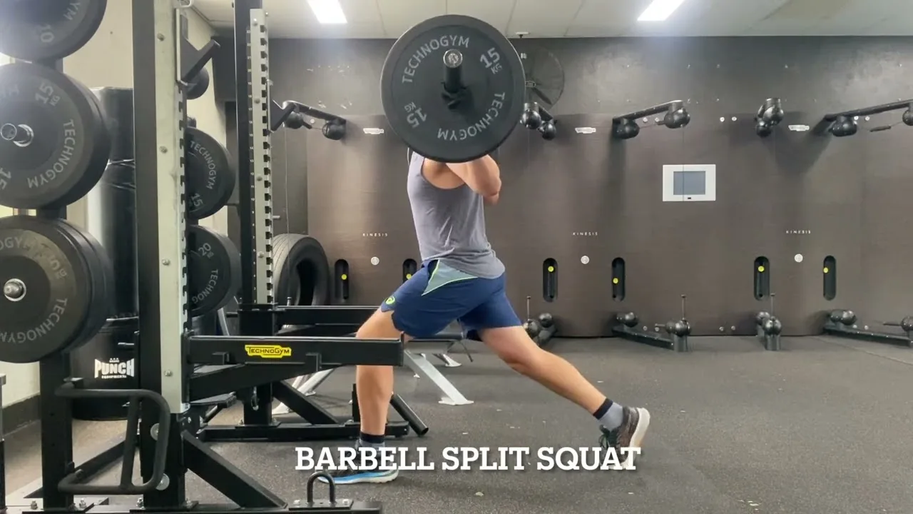
Split Squat vs Squat vs Deadlift: How to tailor your lower body training

The age old question: What’s the best gym activity for my sport? Well, the an…

Unilateral vs Bilateral Training, Part 2: Performance outcomes

In part 1 of this blog, we discussed the generalities associated with bilateral vs unilate…

References

  1. Escamilla RF. Knee biomechanics of the dynamic squat exercise. Med Sci Sports Exerc. 2001;33(1):127-141. https://doi.org/10.1097/00005768-200101000-00020
  2. Young MA, Cook JL, Purdam CR, Kiss ZS, Alfredson H. Eccentric decline squat protocol offers superior results at 12 months compared with traditional eccentric protocol for patellar tendinopathy in volleyball players. Br J Sports Med. 2005;39(2):102-105. https://doi.org/10.1136/bjsm.2003.010587
  3. Hinman RS, Lentzos J, Vicenzino B, Crossley KM. Is patellofemoral osteoarthritis common in middle-aged people with chronic patellofemoral pain? Arthritis Care Res (Hoboken). 2014;66(8):1252-1257. https://doi.org/10.1002/acr.22274
  4. Krommes K, Bjerre A, Thorborg K, Nielsen MF, Hölmich P. Long-term knee health in adults with a history of adolescent Osgood-Schlatter: a national cohort study of patients in secondary care in Denmark 1977-2020. Sports Med. 2025;55(7):1769-1781. https://doi.org/10.1007/s40279-025-02214-5
  5. Yaya-Quezada C, Fanney L, Patel V, Taragin BH, Williams BA, Simoni P, Nguyen JC. Imaging of the pediatric knee. Semin Musculoskelet Radiol. 2024;28(4):462-476. https://doi.org/10.1055/s-0044-1786152
  6. Powers CM. The influence of abnormal hip mechanics on knee injury: a biomechanical perspective. J Orthop Sports Phys Ther. 2010;40(2):42-51. https://doi.org/10.2519/jospt.2010.3337
  7. Macrum E, Bell DR, Boling M, Lewek M, Padua D. Effect of limiting ankle-dorsiflexion range of motion on lower extremity kinematics and muscle-activation patterns during a squat. J Sport Rehabil. 2012;21(2):144-150. https://doi.org/10.1123/jsr.21.2.144
  8. Malliaras P, Cook JL, Kent P. Reduced ankle dorsiflexion range may increase the risk of patellar tendon injury among volleyball players. J Sci Med Sport. 2006;9(4):304-309. https://doi.org/10.1016/j.jsams.2006.03.015
  9. Docking SI, Rio E, Cook J, Carey D, Fortington L. Quantification of Achilles and patellar tendon structure on imaging does not enhance ability to predict self-reported symptoms beyond grey-scale ultrasound and previous history. J Sci Med Sport. 2018;22(2):145-150. https://doi.org/10.1016/j.jsams.2018.07.016

Why Golfer’s Elbow Takes Just as Long to Get Better (And What Actually Works)

Why Golfer’s Elbow Takes Just as Long to Get Better (And What Actually Works)

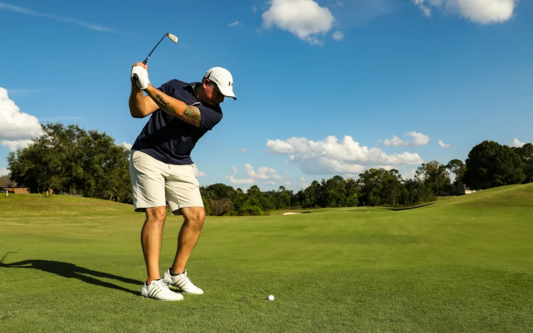
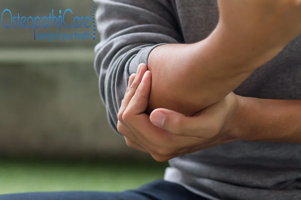
If you’ve had golfer’s elbow (also called medial epicondylalgia), you’ll know it’s not just a “sore elbow.” It’s pain gripping the steering wheel. Pain shaking hands. Pain turning a door handle. Pain gripping a golf club, a cricket bat, or the gym bar. Pain picking up your kids, or even just carrying the shopping in from the car.

And frustratingly, it can hang around for months, sometimes over a year. So why does golfer’s elbow take so long to improve? And are injections like cortisone really the quick fix people hope they are?

Let’s unpack it during this installment of Praxis What You Preach.

Golfer’s elbow is often thought of as tennis elbow’s quieter medial-side cousin. And in a lot of ways, that’s exactly right. If you haven’t already, it’s worth reading our companion piece, Why Tennis Elbow Takes So Long to Get Better. The underlying story of both conditions is remarkably similar. The difference is which side of the elbow, and which tendon, bears the load.

FIRST: IT’S NOT REALLY “INFLAMMATION”

For years golfer’s elbow was called “medial epicondylitis.” The “-itis” was meant to imply inflammation. But when researchers actually biopsied the affected tendon tissue, they didn’t find classic inflammatory cells. What they found was disorganised collagen, immature scar-like tissue, and small blood vessel changes: a picture of degeneration and failed healing, not inflammation (Konarski et al., 2023). That’s why clinicians increasingly prefer the term medial epicondylalgia, or simply call it what it is: a tendinopathy.

The tendon involved is the common flexor-pronator tendon, which attaches to the medial epicondyle (the bony bump on the inside of your elbow). The main offenders are pronator teres and flexor carpi radialis, the muscles that pronate your forearm and flex your wrist. Those are exactly the actions used in a golf swing, a bowling or throwing action, hammering, or repetitive gripping and lifting at work (Konarski et al., 2023). Occupational studies confirm this: forceful gripping and forceful hand-arm exertion are consistently linked with medial epicondylitis, more so than simple repetition alone (Descatha et al., 2003).

This distinction matters clinically. If it were true inflammation, anti-inflammatories and rest would fix it. But because it’s a tendon struggling to remodel and rebuild under load, the fix looks very different, and slower.

WHY IT TAKES SO LONG TO HEAL

Tendon is a tough, relatively low-blood-supply tissue, and it doesn’t rebuild itself quickly. Degenerative tendon changes take time to reverse because the collagen matrix needs to be gradually remodelled through the right amount and type of loading. Too little load and the tendon never adapts; too much, too soon, and you re-irritate it.

This is reflected in how long people tend to have symptoms before they get proper treatment. In one of the key clinical studies on eccentric exercise for golfer’s elbow, patients presenting for treatment had a mean symptom duration of 19 months before starting a structured loading program (Svernlov et al., 2012). Golfer’s elbow is also less common and less studied than tennis elbow, making up roughly 10-20% of all epicondylitis presentations (Wiggins et al., 2018). That partly explains why it’s under-recognised and often mismanaged early, allowed to become chronic before it’s treated properly.

The encouraging part: even patients with long-standing, previously unsuccessfully treated golfer’s elbow can still improve substantially with the right rehabilitation approach. It’s about matching the treatment to what’s actually going on in the tissue (Svernlov et al., 2012).

CORTISONE, PRP, OR LOADING: WHAT DOES THE EVIDENCE ACTUALLY SHOW?

Cortisone injections are still commonly offered for golfer’s elbow, and they do work, in the short term. A direct trial comparing corticosteroid injection with platelet-rich plasma (PRP) in golfer’s elbow patients found the steroid group improved faster in the first month (mean VAS pain score dropping from 5.8 to 3.2). By six months, though, the PRP group had pulled clearly ahead on both pain and function (DASH) scores, with the steroid group’s improvement plateauing at a comparatively higher pain and disability level (Chandrasekaran et al., 2022). In other words: cortisone can quiet things down quickly, but it doesn’t fix the underlying tendon problem, and outcomes can stall or regress.

PRP injections have shown promise for longer-term outcomes in epicondylitis generally, but they’re an adjunct, not a substitute for loading the tendon appropriately. Access, cost, and evidence quality still vary (Konarski et al., 2023).

Meanwhile, structured eccentric loading, the kind a physiotherapist can prescribe and progress, has produced meaningful, durable reductions in pain and improvements in grip strength in dedicated golfer’s elbow studies. Those benefits are maintained at long-term follow-up (Svernlov et al., 2012). It doesn’t work as fast as a needle. But it addresses the actual problem: a tendon that hasn’t had the chance to rebuild its capacity.

WHAT ACTUALLY WORKS

The most consistent, evidence-backed approach is physiotherapy-directed progressive loading of the flexor-pronator group:

Starting with isometric holds to settle pain and maintain strength without aggravating the tendon. Progressing to slow, controlled eccentric and concentric loading of wrist flexion and forearm pronation. Rebuilding grip strength specifically, since grip deficits are common and closely tied to function. Addressing the whole kinetic chain, shoulder, scapular control, and even trunk rotation for golfers and throwers, rather than treating the elbow in isolation. Gradually reintroducing the golf swing, throw, or gym lift once the tendon can tolerate real load.

We don’t currently have a golfer’s-elbow-specific wrist-flexor loading video on our YouTube channel yet. That’s something we should film, so patients have a clear visual guide to follow at home alongside their program.

The Takeaway

Golfer’s elbow isn’t inflammation. It’s a tendon that has lost capacity and needs to rebuild it. That rebuilding takes months, not days, and there’s no shortcut that bypasses the loading process. Cortisone can help short-term flare-ups, but it isn’t a fix. PRP may help longer-term outcomes as an adjunct. The most reliable path back to pain-free gripping, swinging, and lifting is a physiotherapy-guided progressive loading program tailored to your tendon, your sport, and your goals.

Book an appointment at one of our Brisbane clinics today and start moving forward with a clear plan.

Until next time, Praxis What You Preach…

📍 Clinics in Teneriffe, Woolloongabba, and Carseldine

💪 Trusted by athletes. Backed by evidence. Here for everyone.

Related Articles

Why Tennis Elbow Takes So Long to Get Better (And What Actually Works)

Tennis elbow is notoriously slow to heal. Here’s why, and what the evidence actually says helps it along.

Rotator Cuff Repair: A Physiotherapy Guide on Recovery and Rehabilitation

What to expect at every phase of rehab after rotator cuff repair surgery, from protection through to return to sport.

Shoulder Stabilisation and Bankart Repair: Your Path Back to Sport

Recovering from a shoulder stabilisation surgery, particularly a Bankart repair, can feel like a long road back.
References

Chandrasekaran N, Manokaran SV, Jayakaran HB, Ganesh GS. Comparison Between Steroid and PRP Injections in Medial Epicondylitis (Golfer’s Elbow) Patients. International Journal of Pharmaceutical Sciences and Research. 2022;13(12):5139-5144.

Descatha A, Leclerc A, Chastang JF, Roquelaure Y. Medial epicondylitis in occupational settings: prevalence, incidence and associated risk factors. Journal of Occupational and Environmental Medicine. 2003;45(9):993-1001.

Konarski W, Pobozy T, Pobozy K, Domanska J, Konarska K. Current concepts of natural course and in management of medial epicondylitis: a clinical overview. Orthopedic Reviews. 2023;15:84275.

Svernlov B, Hultgren E, Adolfsson L. Medial epicondylalgia (golfer’s elbow) treated by eccentric exercise. Shoulder & Elbow. 2012;4(1):50-55.

Wiggins AJ, Cancienne JM, Camp CL, Degen RM, Altchek DW, Dines JS, Werner BC. Disease Burden of Medial Epicondylitis in the USA Is Increasing: An Analysis of 19,856 Patients From 2007 to 2014. HSS Journal. 2018;14(3):233-237.