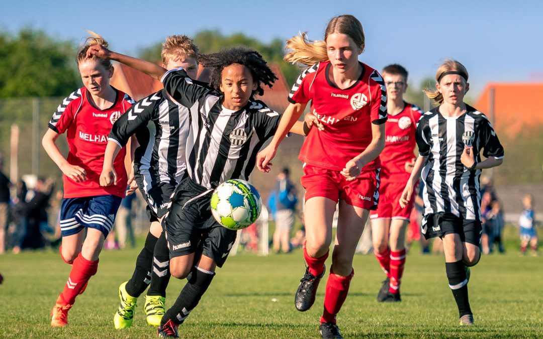
Osgood-Schlatter disease (OSD) (or tibial tuberosity traction apophysitis) is a common condition that affects the knee, primarily in adolescents and young athletes. OSD is more frequently experienced in males 12-15 years old who are involved in activities that require frequent running, jumping, kicking and decelerating, like football (Bezuglov et al 2022). The condition manifests as pain, swelling, and tenderness just below the knee, where the patellar tendon attaches to the tibial tuberosity. Discomfort and potential disruption of daily activities and sports participation is often the result.
A prerequisite for this condition is high loading. The repetitive stress placed on this area during physical activities leads to microtrauma and inflammation, causing symptoms. While the condition is generally self-limiting and tends to resolve as the affected individual completes the growth spurt, physiotherapy plays a pivotal role in effectively managing symptoms, promoting healing, and aiding in a smooth return to physical activities. Various conservative approaches have been studied and recommended in the scientific literature to manage symptoms and aid in the healing process. Interestingly, the condition is strongly associated with Sever’s disease, another growth and loading related injury associated with active young people (Schultz et al 2022). Read on for a general overview of the treatment options supported by scientific research.
Rest and Activity Modification
Rest is often a key component of initial treatment. Reducing or modifying activities that aggravate symptoms, such as avoiding high-impact sports or exercises, can help alleviate strain on the affected area and promote healing. According to a study published in the “Journal of Pediatric Orthopaedics,” activity modification was found to be an effective strategy in managing Osgood-Schlatter Disease, with a significant reduction in pain reported by participants who adhered to activity restrictions.
Physical Therapy and Stretching Exercises
Physical therapy plays a vital role in managing Osgood-Schlatter Disease. A study published in the “Journal of Orthopaedic & Sports Physical Therapy” emphasized the importance of a structured physical therapy program involving stretching exercises for the quadriceps, hamstrings, and calf muscles. These exercises aim to improve muscle flexibility, reduce tension around the knee, and address any muscle imbalances that might contribute to the condition.
Strengthening Exercises
Strengthening exercises focused on the quadriceps and surrounding muscles can help improve biomechanics and stabilize the knee joint. Research published in the American Journal of Sports Medicine highlighted the positive effects of a quadriceps-strengthening program in reducing pain and improving function in individuals with Osgood-Schlatter Disease.
Ice Therapy
Cold therapy, such as applying ice to the affected area, can help reduce inflammation and provide pain relief. A study published in the “Journal of Orthopaedic & Sports Physical Therapy” suggested that ice therapy can be beneficial when used in combination with other conservative treatments.
Non-Steroidal Anti-Inflammatory Drugs (NSAIDs)
NSAIDs, such as ibuprofen, are commonly used to manage pain and inflammation associated with Osgood-Schlatter Disease. However, their use should be supervised by a healthcare professional such as your GP or pharmacist, and long-term or excessive use should be avoided.
Bracing and Taping
Some studies have explored the use of knee braces or taping techniques to offload the patellar tendon and reduce strain on the tibial tuberosity. While research on this aspect is limited, these approaches might offer temporary relief during activities. This can be trial and error as to which technique works best however compression over the tibial tuberosity seems to be the most common strategy.
Education and Activity Guidance
Educating patients and their parents about the condition, its natural history, and appropriate activity modification is crucial. A study in the “Journal of Pediatric Orthopaedics” emphasized the significance of patient education in improving adherence to treatment recommendations and facilitating symptom management.
It’s important to note that each individual’s response to treatment can vary, and a tailored approach is often necessary. In cases where conservative treatments do not provide sufficient relief, and severe pain or functional limitations persist, consultation with a Sports Physician or Orthopaedic surgeon may be warranted. Surgical intervention is rarely indicated and is typically considered only when symptoms are severe, long-lasting, and significantly affecting an individual’s quality of life.
In summary, Osgood-Schlatter Disease can pose significant challenges for adolescents and young athletes, affecting their quality of life and participation in sports. While the condition typically resolves with time and growth plate maturation, the discomfort and limitations it presents can be effectively managed and alleviated with the help of physiotherapy. If you or someone you know is dealing with this condition, get help from our friendly and qualified Praxis physios to individualise an appropriate rehabilitation plan.
Until next time, PREVENT PREPARE PERFORM
Team Praxis
References:
Bezuglov, E., Pirmakhanov, B., Ussatayeva, G., Emanov, A., Valova, Y., Kletsovskiy, A., … & Morgans, R. (2022). The mid-term effect of Osgood-Schlatter disease on knee function in young players from elite soccer academies. ThePhysicianandSportsmedicine, 1-6.
Ciatawi, K., & Dusak, I. W. S. (2022). Osgood-Schlatter disease: A review of current diagnosis and management. CurrentOrthopaedicPractice, 33(3), 294-298.
Schultz, M., Tol, J. L., Veltman, L., & Reurink, G. (2022). Osgood-Schlatter Disease in youth elite football: Minimal time-loss and no association with clinical and ultrasonographic factors. PhysicalTherapyinSport, 55, 98-
Today on the Praxis What We Preach blog, where we shed light on Achilles tendinopathy, a common condition affecting athletes and active individuals. In this article, we will explore the causes, symptoms, and effective treatment strategies for managing Achilles tendinopathy, empowering suffers to return to the things. I draw from personal experience from someone who has had Achilles pain limit my running!
Achilles tendinopathy refers to the degeneration or overload of the Achilles tendon, the band of tissue connecting the calf muscles to the heel bone (calcaneus). This condition primarily affects people engaged in activities involving repetitive jumping, running, or sudden increases in training intensity. Patients with Achilles tendinopathy often experience pain, stiffness, and swelling in the achilles, which can gradually worsen over time. Stiffness and pain is most commonly experienced first thing in the morning, after a long period of sitting or when the achilles has been compressed. Pain can occur in the “mid portion” (pictured below) on in the insertion (as it attaches to the heel bone). This is in an important distinction as these are rehabilitated differently!
Causes and Risks
Achilles tendinopathy typically results from a combination of intrinsic and extrinsic factors. Intrinsic factors include age, reduced flexibility, reduced calf strength / endurance and poor lower limb biomechanics. Extrinsic factors encompass inappropriate footwear, training errors (such as a spike or change in workload), and inadequate warm-up or cool-down routines. Additionally, individuals with systemic conditions like diabetes or rheumatoid arthritis may be more prone to developing Achilles tendinopathy. Understanding these factors is crucial for tailoring treatment plans to address the root causes and minimize the risk of recurrence. But in the most reductionist of terms, Achilles tendinopathy develops due in large part due to a mismatch between loading and the capacity of the tissue.
Diagnosis and Assessment
Accurate diagnosis of Achilles tendinopathy relies on a thorough clinical examination and patient history. Physiotherapists employ various assessment techniques, such as palpation, functional tests, and imaging modalities like ultrasound or MRI, to evaluate the severity and extent of the condition. A self administered questionnaire (VISA-A) can help evaluate symptoms and their effect on physical activity and in turn, the clinical severity. This comprehensive assessment helps determine the appropriate treatment approach, including targeted exercise programs, manual therapy, and other interventions.
Treatment Strategies
Physiotherapy plays a pivotal role in the management of Achilles tendinopathy. Treatment strategies focus on reducing pain, promoting healing, and improving function. These will include calf strengthening exercises, stretching routines and activity modification as frontline options. Moreover, physiotherapists can guide patients in proper footwear selection, gait retraining, and implementing preventive measures to minimize the risk of reinjury.
Rehabilitation and Prevention
Rehabilitation programs are essential for individuals recovering from Achilles tendinopathy. Gradual progression of exercise intensity, functional training, and sport-specific drills enable patients to regain strength, flexibility, and proprioception while minimizing the risk of relapse. Educating patients on proper warm-up and cool-down routines, appropriate footwear selection, and regular monitoring of training loads can significantly contribute to preventing Achilles tendinopathy in the future. One of the common errors patients make is making rehabilitation too easy, or returning to sport too quickly. Again, physiotherapy play a pivotal role in ensuring you undertake a graduated return to loading as the application of mechanical stress to the Achilles tendon promotes tendon healing and remodeling.
Conclusion
Achilles tendinopathy requires a comprehensive approach for effective management. As physiotherapists, our knowledge and expertise are invaluable in helping you overcome this condition and return to their active lifestyles. To discuss your Achilles issues with us to get you back to what you love doing, book online with Praxis today.
Patellofemoral Pain Syndrome (PFPS) is a common condition that affects the knee joint, particularly the area where the kneecap (patella) meets the thigh bone (femur). It is a prevalent issue among athletes, active individuals, and people with certain anatomical factors. In this Praxis What You Preach article, we will explore PFPS, its causes, symptoms, and available treatment options, shedding light on how physiotherapy can effectively manage and alleviate this condition.
What is PFPS?
Patellofemoral Pain Syndrome, also known as runner’s knee or anterior knee pain, occurs when the patella fails to glide smoothly over the femoral groove during knee movement. This causes irritation and inflammation in the patellofemoral joint, specifically the underlying bone, leading to pain, discomfort, loss of function and even swelling. PFPS can be triggered by multiple factors, such as overuse, muscle imbalances, poor biomechanics, weak hip and thigh muscles, improper footwear, and previous knee injuries. Essentially though it is the kneecap joints’ in ability to tolerate the load of the activities being undertaken.
Symptoms and Diagnosis
Common symptoms of PFPS include pain around or behind the patella, especially during activities that involve knee squatting, lunging, bending, climbing / descending stairs, or sitting for extended periods with knees bent (commonly called movie goers knee). These typically can occur when workloads have increased with activities such as running, cycling or weightlifting. Patients may also experience swelling, grinding or even stabbing sensations, and occasionally a feeling of knee instability. A physiotherapist will perform a comprehensive evaluation, considering the patient’s medical history, conducting a physical examination, and possibly using imaging tests, to accurately diagnose PFPS and rule out other potential causes of knee pain.
Treatment and Management
Physiotherapy plays a crucial role in managing and treating PFPS. The primary goal of physiotherapy is to exclude differential diagnoses, alleviate pain, improve knee function, manage aggravating workloads and prevent the recurrence of symptoms. Treatment plans are tailored after a comprehensive history taking and examination to the individual’s specific needs and should include the following components:
Pain Management: Initially, pain and inflammation may be managed through ice therapy, massage, stretching and non-steroidal anti-inflammatory drugs (NSAIDs).
Strengthening Exercises: Targeted exercises aim to strengthen the hip, thigh, and trunk muscles, which can help correct muscle imbalances and improve knee alignment and load tolerance.
Stretching and Flexibility: Stretching exercises can help improve flexibility in the muscles surrounding the knee joint, reducing strain on the patellofemoral joint.
Biomechanical Analysis: A physiotherapist may evaluate the patient’s movement patterns during functional activities such as jumping and running to identify any obvious faulty mechanics that contribute to PFPS. Corrective techniques, gait retraining may be employed.
Activity Modification and Rehabilitation: A gradual return to activities while maintaining a balance between rest and exercise is important to ensure proper healing and prevent re-injury.
Taping: taping has been shown to acutely help reduce symptoms by aiding in the improvement of kneecap tracking through the femoral trochlea (groove where the kneecap runs)
Prevention Strategies
To prevent the onset or recurrence of PFPS, individuals can incorporate the following strategies:
Regular strength and conditioning exercises to maintain muscle balance and strength of the lower limbs and trunk musculature.
Proper warm-up and cool-down routines before and after physical activities.
Gradual progression of activity levels and intensities to avoid overuse injuries.
Being aware of the early signs and symptoms and addressing them promptly.
Is my knee pain osteoarthritis?
In short, No. Patellofemoral Pain Syndrome (PFPS) is not the same as Patellofemoral Joint (PFJ) Osteoarthritis (OA). While both conditions involve the patellofemoral joint, they are distinct entities with different causes and characteristics. As mentioned, PFPS primarily involves pain and dysfunction in the patellofemoral joint, often caused by factors such as overuse, muscle imbalances, or poor biomechanics. It is commonly seen in younger athletes and active individuals. PFPS is characterized by pain around or behind the patella, especially during activities that involve knee bending or loading such as running.
On the other hand, PFJ OA refers to the degeneration and wearing down of the cartilage within the patellofemoral joint. This condition typically occurs in older individuals and is more common in those with a history of knee injuries or conditions such as patellar instability. The primary symptom of patellofemoral joint osteoarthritis is joint pain, stiffness, and swelling, which worsen over time. This pain can be at rest.
While both conditions can cause knee pain and affect the patellofemoral joint, the underlying mechanisms and treatment approaches differ. Physiotherapy plays a crucial role in managing both conditions, but the specific treatment plans and exercises may vary based on the individual’s diagnosis, symptoms, and physical examination findings.
In summary, Patellofemoral Pain Syndrome is a common knee condition that can significantly impact an individual’s daily activities. With a comprehensive physiotherapy approach involving pain management, strengthening exercises, and biomechanical analysis, PFPS can be effectively managed and treated, allowing individuals to regain pain-free movement and engage in their desired activities. If your knee cap pain prevents you from doing the things you want to do, book in with of our expert Praxis team members to discuss getting you back to function!
Until next time, Praxis What You Preach
📍 Clinics in Teneriffe, Buranda, and Carseldine
💪 Trusted by athletes. Backed by evidence. Here for everyone.
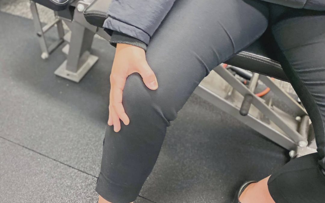
Recently, in an effort to keep the ballooning effects of the all-you-can-eat buffet at bay during my Cricket Australia Indian tour, I ramped up my high-intensity running load. Things were going splendidly — four days of high-intensity running under my belt — until day five, when 90% of the way through a very intense interval session, I tore my hamstring.
I felt the tell-tale sensation so many of my patients describe: a sharp tearing and retraction sensation in my outer thigh while sprinting. I had to pull up immediately and iced the injury straight away. You’ll be happy to hear that I’ve since fully recovered. No longer ‘gun shy’ at my top speeds (which, admittedly, are not that fast!), my strength has vastly improved, and I’m back running at full capacity.
Having treated countless hamstring injuries through my long involvement in recreational, semi-elite, and elite sport — especially with Cricket Australia teams and the Aspley Hornets NEAFL squad — this experience gave me even deeper appreciation for how tricky these injuries can be. Hamstring strains are one of the most common injuries in running athletes, responsible for significant downtime and lost performance. Hamstring injuries have remained the most prevalent injury in professional AFL for the past 21 consecutive seasons (Orchard et al., 2013), with the average 2012 injury costing clubs over $40,000 per player!
Understanding Hamstring Injury Mechanisms
Most hamstring tears occur during the late-swing phase of running, where the hamstring undergoes rapid lengthening while producing high forces (Danielsson et al., 2020). Key risk factors include:
High eccentric loading demands.
Poor neuromuscular control.
Muscle imbalances (particularly hamstrings vs quadriceps).
Fatigue — as evidenced by my own injury, occurring late in a demanding session!
Importantly, the long head of biceps femoris is the most commonly injured muscle, partly due to its higher proportion of fast-twitch fibers and its anatomical position under stretch during running (Martin et al., 2022).
Fatigue, poor trunk/pelvic control, and sudden spikes in high-speed running are emerging as significant contributors to hamstring strain risk, particularly in field and court sports (Martin et al., 2022).
Preventing Hamstring Injuries
The good news is, hamstring injuries can often be prevented with smart training. Strengthening the hamstrings through eccentric exercises like Nordic hamstring curls and single-leg Romanian deadlifts has been shown to reduce injury rates significantly (Al Attar et al., 2017; Martin et al., 2022).
Effective prevention programs should also include:
Agility and trunk stabilization exercises — not just strength work (Martin et al., 2022).
Warm-up routines with dynamic stretching and sport-specific drills.
Monitoring high-speed running loads to avoid sudden spikes in intensity.
Addressing muscle imbalances is key too. Maintaining a healthy strength ratio between the quadriceps and hamstrings — and ensuring good trunk and gluteal control — promotes optimal biomechanics and reduces injury risk (Martin et al., 2022).
Recovering Well After a Hamstring Injury
A proper recovery should include:
Early management: Controlling swelling and pain with ice and appropriate activity modification.
Progressive eccentric strengthening: Integrated carefully to build resilience.
Functional rehabilitation: Sprinting drills, agility work, and sport-specific movements are crucial before returning to full play (Martin et al., 2022).
Interestingly, studies show athletes who follow programs that include eccentric training and trunk stability work have lower reinjury rates than those who just focus on basic strength and stretching (de Visser et al., 2012; Martin et al., 2022).
Return-to-play decisions should be made carefully. Factors like strength symmetry, absence of pain, and readiness for high-speed running should all be considered to reduce the risk of reinjury, which can be as high as 30% otherwise (Martin et al., 2022).
Final Thoughts
Even as a physio, my personal hamstring tear was a stark reminder that fatigue, progressive loading, and structured rehab are vital ingredients for both prevention and recovery. Whether you’re a weekend warrior, a professional cricketer, or just trying to beat the buffet, hamstring health is crucial.
If you’d like help strengthening your hamstrings, managing an existing injury, or optimising your running and performance, feel free to reach out. I (and my hamstrings) would be happy to help!
Till next time, Praxis what you Preach!
Backed by evidence. Trusted by athletes. Here for every body.
References
Al Attar, W.S.A., et al. (2017). The effectiveness of injury prevention programs in reducing the incidence of hamstring injuries in soccer players: a systematic review and meta-analysis. Journal of Physiotherapy, 63(1), 11–17.
Danielsson, B., et al. (2020). Mechanisms of hamstring strain injury: current concepts. Sports Medicine, 50(4), 669–682.
Martin, R.L., et al. (2022). Hamstring strain injury in athletes: Clinical Practice Guidelines. Journal of Orthopaedic & Sports Physical Therapy, 52(3), CPG1–CPG44.
Orchard, J.W., et al. (2013). AFL Injury Report 2012.
Cricket Side Strains in Fast Bowlers: Causes, Prevention, and Rehabilitation
Cricket, a sport demanding significant physical prowess—especially from fast bowlers—often sees athletes pushing their bodies to the limit. This intensity, while integral to performance, can lead to injuries, with side strains being notably common. This Praxis What You Preach blog explores their causes, prevention, and rehabilitation strategies, emphasising the pivotal role of physiotherapy in recovery and return to play.
What is a Side Strain?
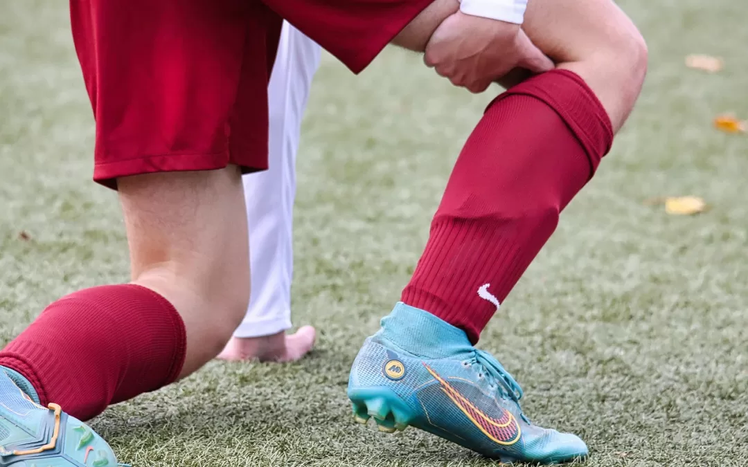
A side strain, or intercostal muscle strain, involves damage to the muscles between the ribs—most commonly the internal obliques. These muscles stabilise the rib cage and generate trunk rotation during bowling. Side strains typically cause acute lateral trunk pain, often on the opposite side to the bowling arm, and usually occur during a forceful delivery, particularly when the front arm pulls down (Orchard et al., 2016). As someone who has torn his side twice bowling, it felt like someone had stabbed me!
Risk Factors
Common risk factors for side strains in fast bowlers include:
Early Season Exposure: Injuries often spike at the beginning of the season due to sudden increases in load (Orchard et al., 2016). This is especially true in athletes transitioning to senior cricket.
Incomplete Rehabilitation from Previous Injuries: Athletes with prior injuries, especially involving the trunk or spine, are more vulnerable (Kountouris & Connell, 2013).
Fast Bowling Biomechanics: Fast bowlers are at significantly greater risk than spinners due to higher trunk rotation velocity and forces (Hardcastle et al., 2015).
Recurrent Injury Rates: There is a high recurrence rate of side strains within the same season if not managed properly (Kountouris & Connell, 2013).
Diagnosis
Diagnosis is primarily clinical, based on sudden pain over the lower ribs, tenderness, and pain during oblique muscle activation (e.g., resisted side bending). Our deep exposure to professional cricket—working closely with elite fast bowlers—gives us a unique edge in accurately diagnosing side strains, often identifying subtle movement patterns and tissue responses that general assessments may overlook. MRI is often used to confirm the diagnosis and assess tear severity (Orchard et al., 2016).
Management and Rehabilitation
Effective rehab balances protection and progressive loading. Key strategies include:
Initial Rest and Pain Management: Reduces inflammation and prevents worsening.
Graduated Physiotherapy: Builds strength and flexibility in core and trunk muscles (Solomen et al., 2015).
Sport-Specific Drills: Return to bowling should include load monitoring and video-based feedback.
Cross-Training: Maintains cardiovascular fitness and lower-body strength without aggravating the injury.
Mild cases resolve in 4–5 weeks; severe injuries may need 6–8 weeks or longer (Orchard et al., 2016).
Prevention Through Strength & Conditioning
A 2015 study by Solomen et al. showed that preseason core strengthening exercises significantly reduced side strain incidence among medium-pace bowlers, highlighting the importance of structured prevention programs (Solomen et al., 2015).
Final Word
In summary, fast bowling is tough work so your body has to be strong and resilient. Good structured strength training helps! If you do suffer a side strain, patience, adherence to rehabilitation protocols, and close monitoring are essential for a successful recovery from a side strain.
If you’re a cricketer struggling with side strains or looking to prevent them, don’t wait for the season to take its toll. At Praxis, we specialise in cricket-specific physiotherapy, grounded in professional-level expertise. With years of hands-on experience in elite cricket environments, we offer advanced assessment, rehab, and return-to-play strategies tailored to the demands of fast bowling. Book a consultation today and let’s get you back to doing what you love—stronger, safer, and better prepared.
The age old question: What’s the best gym activity for my sport? Well – the answer should always be “it depends”. Even the same athlete playing the same sport will have different requirements at different parts of a season. Generally speaking, there are some common exercises in utilised by strength coaches when programming for athletes. The split squat, squat, and deadlift are all compound exercises that target various muscle groups and are commonly included in strength training programs. In today’s Praxis What You Preach article, we are going to breakdown the kinematic (joint angles) and inverse dynamic (joint forces from assumed joint angles) differences between these exercises. We’ll also briefly discuss what sports may benefit, but as just mentioned, the answer is “it depends”.
The Split Squat
The split squat is a unilateral lower body exercise that primarily targets the quadriceps, hamstrings, glutes, and hip stabilisers. It is a personal favourite of mine as I believe it replicates many athletic positions and helps identify any asymmetries there may be. In this exercise, you start in a staggered stance with one foot forward and the other foot positioned behind. The front leg performs most of the work, while the back leg provides support.
The Movement
The front knee flexes and extends, moving vertically.
The rear leg remains relatively stationary, providing balance and stability.
The hip joint of the front leg moves through flexion and extension.
What’s working?
The front leg experiences greater joint forces and moments due to supporting most of the load.
The knee extensors (quadriceps) and hip extensors (glutes) generate the majority of the force to extend the knee and hip joints.
The rear leg primarily acts as a stabilizer rather than generating significant force.
Sports?
The split squat is a versatile exercise that can benefit individuals participating in a wide range of sports. Running, jumping and change of direction field sports such as AFL and soccer seem to benefit well due to the asymmetrical load on the pelvis. The increased loading of the hip stabilising muscles make this a useful exercise for tennis players, volleyballers and track and field (eg triple jumpers) athletes as well.
The Squat
An absolute staple of the gym! The squat is a bilateral lower body exercise that primarily targets the quadriceps, hamstrings, glutes, and lower back muscles. For the sake of this argument, talking about a Barbell back squat. It involves descending into a squatting position while maintaining a relatively upright trunk and then returning to a standing position.
The Movement
The hips and knees flex simultaneously, moving in a coordinated manner.
The knees move forward, tracking over the toes
The torso tilts forward slightly, maintaining a neutral spine but a bit of flex here is fine (and biomechanical studies show you can’t not flex the spine)
What’s Working?
The quadriceps, hamstrings, and glutes generate force to extend the hips and knees during the ascent phase.
The erector spinae and other lower back muscles provide stabilization and contribute to maintaining an upright posture.
The knee extensors (quadriceps) experience higher forces and moments during the descent and ascent phases.
Sports?
Squats help with vertical force generation so jumping sports like basketball and volleyball are sports that would benefit. The Barbell back squat is also central in powerlifting, olympic lifting and Crossfit. Given you can load significant weights to the bar, back squats are also useful for football codes whe are required to absorb impacts during tackles.
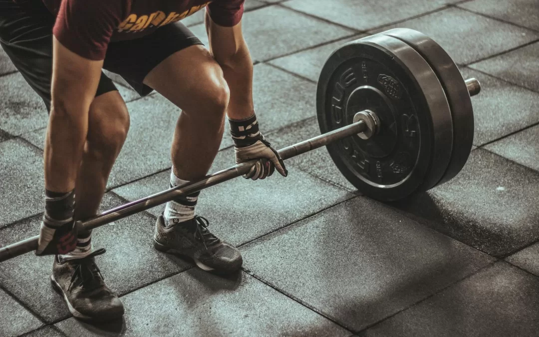
The Deadlift
The deadlift is a bilateral exercise that primarily targets the posterior chain, including the glutes, hamstrings, erector spinae, and upper back muscles. It involves lifting a loaded barbell or other weight from the floor while maintaining proper form.
The Movement
The hips hinge backward, allowing the torso to lean forward while maintaining a neutral spine.
The knees flex to a lesser extent compared to the squat.
The barbell moves vertically in a straight line close to the body.
What’s Working?
The glutes, hamstrings, and erector spinae generate force to extend the hips and maintain a neutral spine.
The quadriceps contribute to knee extension.
The upper back muscles help stabilize the spine and prevent excessive forward flexion.
The lower back muscles experience significant forces and moments due to their role in maintaining spinal alignment.
Sports?
Powerlifting, Olympic lifting and Crossfit are the obvious ones that spring to mind. But tackling sports such as rugby can benefit. Given the predominance of back musculature, rowers will benefit here. Wrestlers and MMA athletes will also benefit due to the whole body nature of a deadlift.
Overall, while all three exercises involve lower body movements, they differ in terms of joint angles, muscle activation patterns, and force distribution. Understanding these differences can help tailor training programs to specific goals and individual needs. We also modify these exercises further to tailor our rehabilitation needs, In that vein, it’s important to be conscious of technique when performing these exercises to maximise their effectiveness and reduce the risk of injury.
So if you are growing stale in your lower body workouts, try and mix it up with some of the above. There are also plenty of variations of the above to alter the movement and forces even more! If you are after some help to modify your gym program, chat to us today – we are here to help!
Until next time, Praxis What You Preach…
📍 Clinics in Teneriffe, Buranda, and Carseldine
💪 Trusted by athletes. Backed by evidence. Here for everyone.