ACL Rehabilitation: The Role of Physiotherapy in Returning to Life, Activity, and Sport

ACL Rehabilitation: The Role of Physiotherapy in Returning to Life, Activity, and Sport

On today’s Praxis what you Preach, we cover a very common injury here in Australia – the Anterior Cruciate ligament (ACL) injury. At Praxis Physiotherapy, we understand that recovering from ACL reconstruction is more than just healing a knee — it’s about restoring confidence, movement, and returning to the activities and lifestyle that matter most to each person. Physiotherapists are uniquely placed to guide this journey from surgery through to return to everyday function, recreation, and sport.

What is an ACL Rupture?

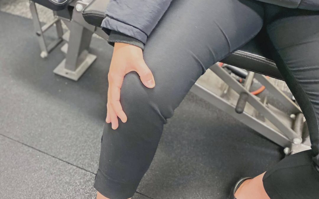
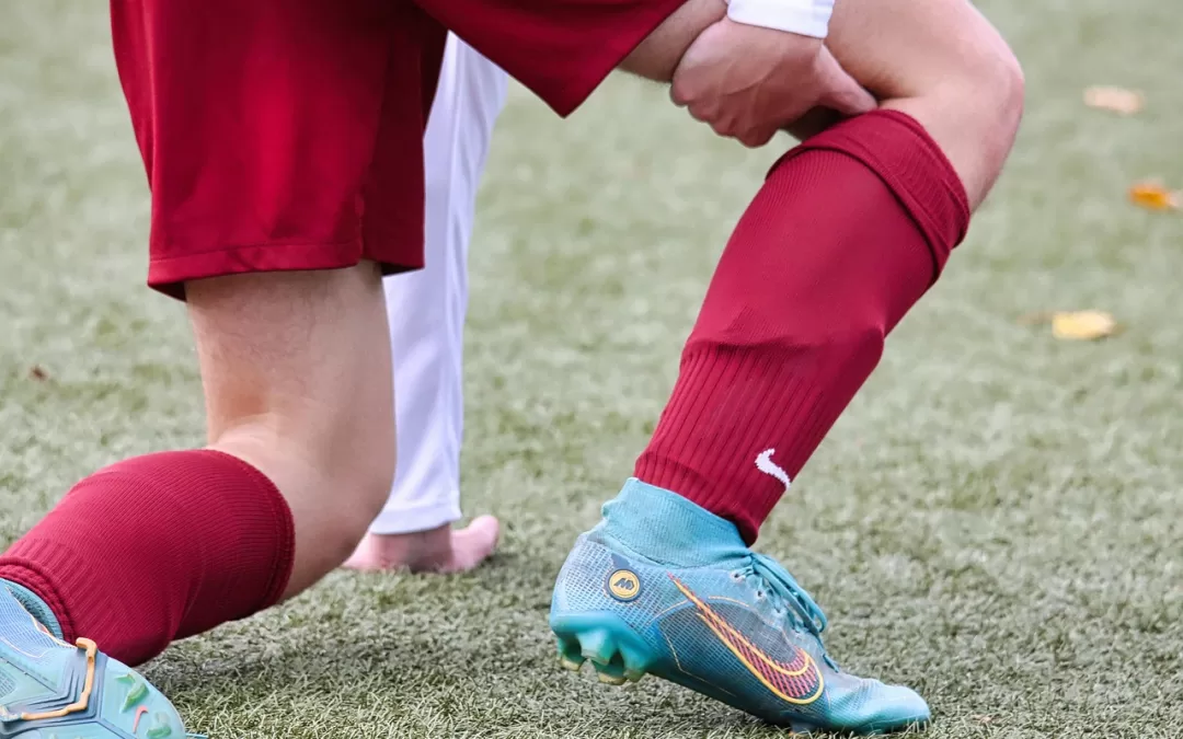
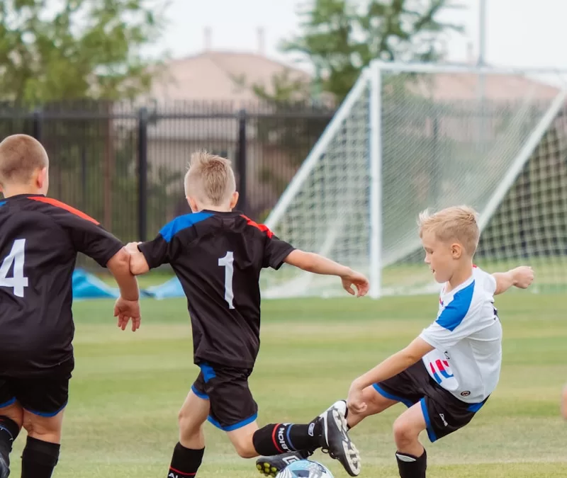
The ACL is one of the key stabilising ligaments of the knee, crucial for controlling rotation and forward movement of the tibia. An ACL rupture typically occurs during sudden changes in direction, pivoting, or awkward landings — common in sports like AFL, soccer, basketball, and netball. It most often affects young, active individuals, particularly females, due to biomechanical and hormonal factors. While not all ACL injuries require surgery, those with complete ruptures who wish to return to cutting or pivoting sports usually undergo ACL reconstruction. Regardless of the surgical decision, structured rehabilitation guided by a physiotherapist is essential for a successful recovery and long-term knee health.

The Importance of Physiotherapy in ACL Rehab

Research shows that while around 80% of individuals return to some form of sport after ACL reconstruction, only 65% return to their preinjury level and just 55% to competitive levels (Andrade et al. 2020). Physiotherapy plays a vital role in improving these outcomes by guiding progressive rehabilitation and using structured criteria-based progressions.

Physiotherapy-led rehabilitation should begin early, with emphasis on knee mobilisation, weight-bearing as tolerated, and initiation of neuromuscular training (Andrade et al. 2020). The BJSM systematic review of clinical guidelines for ACL rehab supports early kinetic chain exercises (both open and closed), strength training, cryotherapy, and neuromuscular stimulation when indicated (Andrade et al. 2020).

From Healing to Performance: A Continuum

Recovery after ACL surgery should follow a continuum from impairment-based care to performance restoration. This includes early pain and swelling control, progressive strength and range of motion restoration, motor control retraining, and sport-specific preparation. At Praxis, we follow a phase-based rehabilitation model tailored to individual needs. These needs may include the type of surgical graft used, concurrent injury (e.g meniscus / MCL), the operating surgeon’s post-op protocols, the patient’s goals, sport-specific demands, timelines for return to competition, and previous levels of function — all of which require thoughtful and collaborative clinical decision-making.

Unfortunately, studies show that many patients are discharged before meeting strength or performance benchmarks — particularly in strength-focused exercises like the split squat, squat, or deadlift, which play a vital role in ACL rehab progression. For example, performing around 22 single-leg sit-to-stands is one such late-stage benchmark that reflects adequate quadriceps strength and control before return to sport (Welling et al 2018). Nichols et al. (2021) found that most published rehabilitation protocols emphasize endurance and hypertrophy without progressing to the strength or power needed to reduce reinjury risk. This underlines the need for physiotherapists to include high-intensity, sports specific strength training and late-stage performance metrics as patients near return to sport.

Addressing Muscle Atrophy and Weakness

Quadriceps atrophy remains a key barrier to recovery post-ACL reconstruction. Evidence supports adjunct interventions such as neuromuscular electrical stimulation and blood flow restriction (BFR) training to combat muscle loss, particularly in the early post-operative period (Charles et al. 2020). BFR combined with low-load resistance exercise has been shown to reduce muscle wasting and promote strength gains when higher loads are contraindicated — we explore this more in our Blood Flow Restriction Training blog. We use this frequently at Praxis Physiotherapy in both reformer pilates and early gym based settings. 

The Role of the Physio: More Than Just Exercise

Our job as physiotherapists goes beyond prescribing exercises. We support patients through the emotional and motivational challenges of recovery, address fear of re-injury, and help them develop the confidence to return to sport or physically demanding jobs. We tailor plans based on functional goals, sport-specific needs, and personal circumstances.

At Praxis, this also means working closely with coaches, GPs, surgeons, and families to ensure clear communication and aligned expectations. For sporting patients, this might include on-field rehab or comprehensive return-to-play assessments in collaboration with clubs and teams.

A Collaborative, High-Performance Rehabilitation Environment

At Praxis Physiotherapy, we bring high-performance rehab principles to all patients — not just elite athletes. Our team has provided physiotherapy services to the Aspley Hornets AFL Club since 2014, giving us deep insight into the physical and mental demands of competitive sport. We apply this same standard of care to everyday athletes, weekend warriors, and anyone seeking to return to an active lifestyle.

We also work closely with orthopaedic knee and shoulder surgeon Dr. Kelly Macgroarty, including in-room triage consulting, ensuring a seamlessly integrated, evidence-informed rehabilitation pathway. This collaboration allows us to align surgical timelines, post-op considerations, and physiotherapy progressions — from day one to return to sport.

Our clinical culture is shaped by exposure to elite-level sports environments, including AFL, representative athletics, and professional national cricket programs. But rather than highlight individual accolades, we’re most proud of the high clinical standards and systems-based approach that ensure our entire team delivers the same quality of care — no matter who walks through the door.

Each of our Brisbane based clinics includes access to gym facilities and reformer Pilates equipment, allowing for real-world, function-driven exercise. These resources support patients to not only recover structurally but also return to high levels of strength, coordination, and performance in line with the latest evidence-based guidelines.

A Message to Our Patients

Whether you’re an athlete aiming for competitive return or someone wanting to run after your kids again, we bring the same level of care and attention to your ACL rehab. Recovery is not just about timelines — it’s about building back strength, movement, and trust in your knee. Ready to get started with your own recovery plan? Explore the ACL physiotherapy services at Praxis and book an appointment today.

Until next time, Praxis What You Preach…

📍 Clinics in Teneriffe, Buranda, and Carseldine
💪 Trusted by athletes. Backed by evidence. Here for everyone.

For more insights into long-term knee health, including non-surgical rehab, check out our Knee Osteoarthritis blog.


References

Andrade R, et al. (2020). How should clinicians rehabilitate patients after ACL reconstruction? A systematic review of clinical practice guidelines. Br J Sports Med, 54(9), 512–519.

Kochman M, et al. (2022). ACL Reconstruction: Which Additional Physiotherapy Interventions Improve Early-Stage Rehabilitation? Int J Environ Res Public Health, 19(23), 15893.

Charles D, et al. (2020). A systematic review of the effects of blood flow restriction training on quadriceps muscle atrophy and circumference post ACL reconstruction. Int J Sports Phys Ther, 15(6), 882–889.

Nichols ZW, et al. (2021). Is resistance training intensity adequately prescribed to meet the demands of returning to sport following ACL repair? A systematic review. BMJ Open Sport Exerc Med, 7(1), e001144.

Welling W, Benjaminse A, Gokeler A, Otten E, & Seil R. (2018). Low rates of patients meeting return to sport criteria 9 months after anterior cruciate ligament reconstruction: a prospective longitudinal study. Knee Surg Sports Traumatol Arthrosc, 26(12), 3636–3644.

Capping kneecap pain – Your guide to Anterior Knee Pain (Patellofemoral Pain Syndrome)

Capping kneecap pain – Your guide to Anterior Knee Pain (Patellofemoral Pain Syndrome)

Patellofemoral Pain Syndrome (PFPS) is a common condition that affects the knee joint, particularly the area where the kneecap (patella) meets the thigh bone (femur). It is a prevalent issue among athletes, active individuals, and people with certain anatomical factors. In this Praxis What You Preach article, we will explore PFPS, its causes, symptoms, and available treatment options, shedding light on how physiotherapy can effectively manage and alleviate this condition.

What is PFPS?

Patellofemoral Pain Syndrome, also known as runner’s knee or anterior knee pain, occurs when the patella fails to glide smoothly over the femoral groove during knee movement. This causes irritation and inflammation in the patellofemoral joint, specifically the underlying bone, leading to pain, discomfort, loss of function and even swelling. PFPS can be triggered by multiple factors, such as overuse, muscle imbalances, poor biomechanics, weak hip and thigh muscles, improper footwear, and previous knee injuries. Essentially though it is the kneecap joints’ in ability to tolerate the load of the activities being undertaken.

Symptoms and Diagnosis

Common symptoms of PFPS include pain around or behind the patella, especially during activities that involve knee squatting, lunging, bending, climbing / descending stairs, or sitting for extended periods with knees bent (commonly called movie goers knee). These typically can occur when workloads have increased with activities such as running, cycling or weightlifting. Patients may also experience swelling, grinding or even stabbing sensations, and occasionally a feeling of knee instability. A physiotherapist will perform a comprehensive evaluation, considering the patient’s medical history, conducting a physical examination, and possibly using imaging tests, to accurately diagnose PFPS and rule out other potential causes of knee pain.

Treatment and Management

Physiotherapy plays a crucial role in managing and treating PFPS. The primary goal of physiotherapy is to exclude differential diagnoses, alleviate pain, improve knee function, manage aggravating workloads and prevent the recurrence of symptoms. Treatment plans are tailored after a comprehensive history taking and examination to the individual’s specific needs and should include the following components:

  • Pain Management: Initially, pain and inflammation may be managed through ice therapy, massage, stretching and non-steroidal anti-inflammatory drugs (NSAIDs).
  • Strengthening Exercises: Targeted exercises aim to strengthen the hip, thigh, and trunk muscles, which can help correct muscle imbalances and improve knee alignment and load tolerance.
  • Stretching and Flexibility: Stretching exercises can help improve flexibility in the muscles surrounding the knee joint, reducing strain on the patellofemoral joint.
  • Biomechanical Analysis: A physiotherapist may evaluate the patient’s movement patterns during functional activities such as jumping and running to identify any obvious faulty mechanics that contribute to PFPS. Corrective techniques, gait retraining may be employed.
  • Activity Modification and Rehabilitation: A gradual return to activities while maintaining a balance between rest and exercise is important to ensure proper healing and prevent re-injury.
  • Taping: taping has been shown to acutely help reduce symptoms by aiding in the improvement of kneecap tracking through the femoral trochlea (groove where the kneecap runs)

Prevention Strategies

To prevent the onset or recurrence of PFPS, individuals can incorporate the following strategies:

  • Regular strength and conditioning exercises to maintain muscle balance and strength of the lower limbs and trunk musculature.
  • Proper warm-up and cool-down routines before and after physical activities.
  • Gradual progression of activity levels and intensities to avoid overuse injuries.
  • Being aware of the early signs and symptoms and addressing them promptly.

Is my knee pain osteoarthritis?

In short, No. Patellofemoral Pain Syndrome (PFPS) is not the same as Patellofemoral Joint (PFJ) Osteoarthritis (OA). While both conditions involve the patellofemoral joint, they are distinct entities with different causes and characteristics. As mentioned, PFPS primarily involves pain and dysfunction in the patellofemoral joint, often caused by factors such as overuse, muscle imbalances, or poor biomechanics. It is commonly seen in younger athletes and active individuals. PFPS is characterized by pain around or behind the patella, especially during activities that involve knee bending or loading such as running.

On the other hand, PFJ OA refers to the degeneration and wearing down of the cartilage within the patellofemoral joint. This condition typically occurs in older individuals and is more common in those with a history of knee injuries or conditions such as patellar instability. The primary symptom of patellofemoral joint osteoarthritis is joint pain, stiffness, and swelling, which worsen over time. This pain can be at rest.

While both conditions can cause knee pain and affect the patellofemoral joint, the underlying mechanisms and treatment approaches differ. Physiotherapy plays a crucial role in managing both conditions, but the specific treatment plans and exercises may vary based on the individual’s diagnosis, symptoms, and physical examination findings.

In summary, Patellofemoral Pain Syndrome is a common knee condition that can significantly impact an individual’s daily activities. With a comprehensive physiotherapy approach involving pain management, strengthening exercises, and biomechanical analysis, PFPS can be effectively managed and treated, allowing individuals to regain pain-free movement and engage in their desired activities. If your knee cap pain prevents you from doing the things you want to do, book in with of our expert Praxis team members to discuss getting you back to function!

Until next time,

Praxis What You Preach

Team Praxis

Hamstring Strain Injuries: Lessons from Personal Experience and the Latest Research

Hamstring Strain Injuries: Lessons from Personal Experience and the Latest Research

Recently, in an effort to keep the ballooning effects of the all-you-can-eat buffet at bay during my Cricket Australia Indian tour, I ramped up my high-intensity running load. Things were going splendidly — four days of high-intensity running under my belt — until day five, when 90% of the way through a very intense interval session, I tore my hamstring.

I felt the tell-tale sensation so many of my patients describe: a sharp tearing and retraction sensation in my outer thigh while sprinting. I had to pull up immediately and iced the injury straight away. You’ll be happy to hear that I’ve since fully recovered. No longer ‘gun shy’ at my top speeds (which, admittedly, are not that fast!), my strength has vastly improved, and I’m back running at full capacity.

Having treated countless hamstring injuries through my long involvement in recreational, semi-elite, and elite sport — especially with Cricket Australia teams and the Aspley Hornets NEAFL squad — this experience gave me even deeper appreciation for how tricky these injuries can be. Hamstring strains are one of the most common injuries in running athletes, responsible for significant downtime and lost performance. Hamstring injuries have remained the most prevalent injury in professional AFL for the past 21 consecutive seasons (Orchard et al., 2013), with the average 2012 injury costing clubs over $40,000 per player!

Understanding Hamstring Injury Mechanisms

Most hamstring tears occur during the late-swing phase of running, where the hamstring undergoes rapid lengthening while producing high forces (Danielsson et al., 2020). Key risk factors include:

  • High eccentric loading demands.

  • Poor neuromuscular control.

  • Muscle imbalances (particularly hamstrings vs quadriceps).

  • Fatigue — as evidenced by my own injury, occurring late in a demanding session!

Importantly, the long head of biceps femoris is the most commonly injured muscle, partly due to its higher proportion of fast-twitch fibers and its anatomical position under stretch during running (Martin et al., 2022).

Fatigue, poor trunk/pelvic control, and sudden spikes in high-speed running are emerging as significant contributors to hamstring strain risk, particularly in field and court sports (Martin et al., 2022).

Preventing Hamstring Injuries

The good news is, hamstring injuries can often be prevented with smart training. Strengthening the hamstrings through eccentric exercises like Nordic hamstring curls and single-leg Romanian deadlifts has been shown to reduce injury rates significantly (Al Attar et al., 2017; Martin et al., 2022).

Effective prevention programs should also include:

  • Agility and trunk stabilization exercises — not just strength work (Martin et al., 2022).

  • Warm-up routines with dynamic stretching and sport-specific drills.

  • Monitoring high-speed running loads to avoid sudden spikes in intensity.

Addressing muscle imbalances is key too. Maintaining a healthy strength ratio between the quadriceps and hamstrings — and ensuring good trunk and gluteal control — promotes optimal biomechanics and reduces injury risk (Martin et al., 2022).

Recovering Well After a Hamstring Injury

A proper recovery should include:

  • Early management: Controlling swelling and pain with ice and appropriate activity modification.

  • Progressive eccentric strengthening: Integrated carefully to build resilience.

  • Functional rehabilitation: Sprinting drills, agility work, and sport-specific movements are crucial before returning to full play (Martin et al., 2022).

Interestingly, studies show athletes who follow programs that include eccentric training and trunk stability work have lower reinjury rates than those who just focus on basic strength and stretching (de Visser et al., 2012; Martin et al., 2022).

Return-to-play decisions should be made carefully. Factors like strength symmetry, absence of pain, and readiness for high-speed running should all be considered to reduce the risk of reinjury, which can be as high as 30% otherwise (Martin et al., 2022).

Final Thoughts

Even as a physio, my personal hamstring tear was a stark reminder that fatigue, progressive loading, and structured rehab are vital ingredients for both prevention and recovery. Whether you’re a weekend warrior, a professional cricketer, or just trying to beat the buffet, hamstring health is crucial.

If you’d like help strengthening your hamstrings, managing an existing injury, or optimising your running and performance, feel free to reach out. I (and my hamstrings) would be happy to help!

Till next time, Praxis what you Preach!

Backed by evidence. Trusted by athletes. Here for every body.

References

  • Al Attar, W.S.A., et al. (2017). The effectiveness of injury prevention programs in reducing the incidence of hamstring injuries in soccer players: a systematic review and meta-analysis. Journal of Physiotherapy, 63(1), 11–17.

  • Danielsson, B., et al. (2020). Mechanisms of hamstring strain injury: current concepts. Sports Medicine, 50(4), 669–682.

  • Martin, R.L., et al. (2022). Hamstring strain injury in athletes: Clinical Practice Guidelines. Journal of Orthopaedic & Sports Physical Therapy, 52(3), CPG1–CPG44.

  • Orchard, J.W., et al. (2013). AFL Injury Report 2012.

Plantar Fasciopathy: Understanding how to heal your heel pain

Plantar Fasciopathy: Understanding how to heal your heel pain

Feel like your walking on glass in the mornings?  Those first few steps after a long period of sitting hurt the underside of your heel? Struggling to stand at the end of a long day due to your feet? If so, then you may have plantar fasciopathy, also known as plantar fasciitis. Plantar fasciopathy is a common condition that affects the plantar fascia – a thick band of connective tissue on the bottom of the foot. Plantar fasciopathy commonly affects individuals between the ages of 40 and 60, but can affect almost anyone. In this article, we will delve into the causes, symptoms, treatment options, and preventive measures to help you understand, and more importantly manage, this condition.

Causes and Symptoms

Plantar fasciopathy is often caused by repetitive strain or excessive loading of the plantar fascia, leading to microtears and inflammation. Factors such as overuse, improper footwear, high-impact activities, flat or high-arched feet, and tight calf muscles can contribute to its development. The condition is characterised by sharp pain or a dull ache on the underside of the heel or along the arch of the foot. Pain is typically worse in the morning or after periods of inactivity, and may improve with movement. Standing for long periods or walking on hard floor can also be aggravating.

Treatment Options

The treatment of plantar fasciopathy focuses on reducing pain, promoting load tolerance, and addressing the underlying causes. Physiotherapy interventions play a crucial role in managing this condition. Therapeutic techniques such as manual therapy, stretching exercises, and strengthening exercises can help relieve pain, improve flexibility, and restore foot function. Specifically, improving the windlass mechanism (a phenomena that refers to the tightening of the plantar fascia during the push-off phase of walking or running when you big toe extends). This mechanism helps distribute forces evenly throughout the foot and reduces strain on the plantar fascia. More generally, improvement of the footy intrinsics and plantar flexors more generally have been shown to reduce the severity and duration of symptoms as well.

Additionally, the use of orthotics, taping, or night splints may provide support and alleviate symptoms. Extracorporeal shockwave therapy (ESWT) and ultrasound therapy are also viable treatment options in some cases. In severe or persistent cases, corticosteroid injections or surgery may be considered, though this is usually reserved for when conservative measures have failed.

Preventive Measures

Prevention is key to reducing the risk of plantar fasciopathy starting in the first instance. If you are keen to ‘pound the pavement’ for example, then gradually increase activity levels. Avoid sudden changes in intensity or duration to prevent overloading the foot. This may mean dancing long bouts for the first time in a while, or returning to running post injury. Wear footwear that provides adequate arch support and cushioning. Understand the importance of regular stretching exercises for the calf muscles and plantar fascia.

As physiotherapy professionals, we understand that addressing the symptoms of plantar fasciopathy early is essential for providing effective care. At Praxis, effective care means arming you with adequate advice and education so you can help manage the symptoms yourself. Further, implementing appropriate treatment options and emphasizing preventive measures, we support individuals in overcoming foot pain and restoring quality of life. After all, we aim to Prevent, Prepare, Perform! So if you have heel pain that is stopping you from doing what you would like to do, discuss it with our knowledgeable team today!

Until next time – Praxis What You Preach!

📍 Clinics in Teneriffe, Buranda, and Carseldine

💪 Trusted by athletes. Backed by evidence. Here for everyone.

Understanding Sever’s Disease: A Common Foot Condition in Active Growing Children

Understanding Sever’s Disease: A Common Foot Condition in Active Growing Children

Understanding Sever’s Disease in Growing Children

Sever’s disease — medically known as calcaneal apophysitis — is a common heel condition affecting active children, especially during growth spurts. Despite its name, it’s not a “disease” in the traditional sense, but an overuse injury of the growth plate at the back of the heel.

What Causes Sever’s Disease?

Sever’s disease develops when repetitive stress irritates the growth plate in the heel bone (the calcaneal apophysis). During adolescence, especially between ages 8–15, the heel bone may grow faster than the surrounding muscles and tendons, causing excessive tension at the Achilles insertion site. When coupled with repetitive impact — such as running, jumping, or playing on hard surfaces — this mechanical overload leads to inflammation and pain.

It’s especially common in sports like soccer, basketball, AFL, netball, and gymnastics. Kids going through growth spurts, or who are highly active without sufficient recovery, are most at risk. Tight calf muscles, poor footwear, and biomechanical factors like flat feet or poor shock absorption may also contribute.

Common Symptoms

The main symptom is heel pain that worsens during physical activity and settles with rest. Children may complain of:

  • Pain or tenderness at the back or underside of the heel

  • Limping or toe-walking, particularly after sport

  • Discomfort when pressing on the heel or squeezing it from both sides

  • Stiffness first thing in the morning or after periods of inactivity

Symptoms are usually one-sided but can be bilateral. If left unaddressed, the pain can start to interfere with participation in sport and physical education at school.

Diagnosis

A diagnosis is usually made through clinical history and physical examination by a physiotherapist. Key indicators include heel pain during activity, recent growth, and tenderness at the posterior heel. The “squeeze test” — applying gentle pressure to both sides of the heel — is often positive.

Imaging (X-ray or MRI) is rarely needed unless symptoms persist longer than expected, or there is suspicion of another diagnosis. Importantly, a visible growth plate on X-ray in this age group is normal and not a reason for concern in itself.

Treatment and Management

Treatment is focused on reducing inflammation, offloading the heel, and supporting the child’s return to normal function. It is important to reassure both child and parent that this is a temporary, self-limiting condition.

Key management strategies include:

  • Load modification: Avoiding or reducing high-impact activity is key, especially sports with frequent jumping or sprinting. Your physio can help create a weekly plan to reduce flare-ups while keeping your child engaged and active.

  • Ice: Icing the heel after sport can reduce inflammation and pain, especially in the early stages.

  • Heel lifts or orthotics: Studies, including the 2016 randomised trial by James et al., show that both orthotic devices and cushioned heel lifts can effectively reduce heel stress. These inserts help absorb shock and reduce Achilles tendon tension.

  • Calf stretching and strengthening: Tight calf muscles increase load on the heel. Scharfbillig et al. (2008) emphasised the role of flexibility programs, particularly eccentric calf training, in improving outcomes.

  • Footwear advice: Supportive, well-fitted athletic shoes are essential. Avoid barefoot running or flat-soled footwear during recovery.

  • Manual therapy and taping: In some cases, hands-on techniques and taping methods may be used to reduce load on the Achilles insertion.

According to the trial by Weert et al. (2016), physical therapy combining load management and exercise-based rehab was just as effective as orthotic devices. This supports a flexible treatment approach tailored to the child’s specific needs and activity level.

Medication: Short courses of anti-inflammatory medication such as ibuprofen can help in more severe cases, especially when pain interferes with sleep or daily function. However, these should always be used under medical advice.

Prevention and Long-Term Outlook

The prognosis for Sever’s disease is excellent. Most children recover fully once the growth plate fuses — typically by age 15 for boys and 13 for girls. The condition does not cause permanent damage, though symptom duration can vary from a few weeks to several months depending on activity levels and adherence to management.

To reduce the risk of recurrence:

  • Encourage a proper warm-up and cool-down routine

  • Ensure sport participation is balanced with adequate rest

  • Maintain calf flexibility and foot strength

  • Use shock-absorbing shoes or orthotics during growth spurts

  • Avoid large increases in training volume or intensity

It’s also helpful to educate kids and parents that some discomfort during growth phases is normal, but persistent pain warrants a review. At Praxis Physiotherapy, our clinicians are experienced in managing growing athletes — and ensuring they don’t miss more game time than necessary.

Summary

Sever’s disease is a common and manageable cause of heel pain in growing children. Early recognition, temporary load reduction, and a guided rehab program can ensure a smooth recovery and quick return to sport. If your child is experiencing heel pain that isn’t improving with rest, book in with the friendly and knowlegable team at Praxis Physiotherapy for a tailored management plan.

Until next time, Praxis What You Preach

📍 Clinics in Teneriffe, Buranda, and Carseldine

💪 Trusted by athletes. Backed by evidence. Here for everyone.


References

James, A. M., Williams, C. M., & Haines, T. P. (2016). Effectiveness of footwear and foot orthoses for calcaneal apophysitis: a 12-month factorial randomised trial. British Journal of Sports Medicine, 50(20), 1268–1275. https://doi.org/10.1136/bjsports-2015-094986

Scharfbillig, R. W., Jones, S., & Scutter, S. D. (2008). Sever’s Disease: What Does the Literature Really Tell Us? Journal of the American Podiatric Medical Association, 98(3), 212–223. https://doi.org/10.7547/0980212

Weert, H. C., van Dijk, C. N., & Struijs, P. A. (2016). Treatment of Calcaneal Apophysitis: Wait and See Versus Orthotic Device Versus Physical Therapy. Journal of Pediatric Orthopaedics, 36(2), 152–157. https://doi.org/10.1097/BPO.0000000000000417