Fact or Fiction Friday – Lower back Pain and MRI’s

Fact or Fiction Friday – Lower back Pain and MRI’s

I need to get an MRI to help with the management of my lower back pain

Answer – FICTION

In a recent narrative review, Wang and colleagues (2018) concluded that MRI imaging in the early stages of lower back pain can have detrimental effects including more pain, less improvement, higher risk of surgery and worse overall health status. In fact, one study reported that patients that received an MRI within the first month had an 8x greater risk for surgery and 5x more medical costs!

If you do NOT present with severe neurological deficits, signs of a serious or specific underlying condition or have persistent pain >6 weeks which is unresponsive to conservative treatment then there likely isn’t a need for further investigation!

To get help with your long standing back pain or even that acute flare up, give us a call on (07) 3102 3337 or book online  so we can sort you out.

#praxiswhatyoupreach #praxisphysio #factorfictionfriday #physioeducation #preventprepareperform #pain #backpain #lowerbackpain #MRI #patienteducation

Wang Y, et al. Informed appropriate imaging for low back pain management: A narrative review. Journal of Orthopaedic Translation. 2018.

Stingers AKA Neural traction injuries

Stingers AKA Neural traction injuries

SUMMARY:

  • Stingers are essentially a tractioning of the neural system
  • This can cause pain, movement and sensation changes
  • Chronic traction to the nervous system can have a cumulative effect on nerve function
  • The more damage to the nerve, the more serious the outcome
  • We shouldn’t be as dismissive of “stingers”, particularly if they are recurrent
  • Physiotherapy has a role to play

STINGERS:

Stingers have been the catch cry of many contact sports over the years. Often dismissed as ‘just a stinger’, trauma to the nervous system should probably be taken a little more seriously, as we delve into detail today.

Stingers are most commonly experienced in contact sports whereby the shoulder of a players is forcefully depressed, as experienced with a tackle in NRL or union. A large range of motion over a short period of time can result in a ‘traction’ of the Brachial Plexus (a network of nerves formed from exiting branches of the spinal cord in the neck that transverse to the shoulder and arm). Thus network of nerves sends signals from your spinal cord to your shoulder, arm and hand and thereby providing feeling and movement to these regions.

BACKGROUND:

To understand a stinger injury, a clear understanding of nerves and nerve related injuries is required. A nerve is an enclosed, cable-like bundle of nerve fibres called axons, in the peripheral nervous system. A nerve provides a common pathway for the electrochemical nerve impulses that provide a number of functions, including getting our muscles to move!

Seddon and Sunderland present a five-grade classification scale for nerve related injuries[1-4]. Figure 1 illustrates the physiological changes that occur through each grade of injury. Essentially it outlines the greater amount of disruption to the anatomy of the nerve.

The more damage to the nerve, the more serious the outcome. Table 1 outlines the three different grades of stinger injuries.[4, 5] The most common stinger is a grade 1 injury, which represents a neurapraxia, or nerve stretch injury, without axonal disruption.[4] In an acute setting, this can result in motor and sensory loss/changes, which usually resolve within minutes.[1, 4] Grade 2 and 3 involve a higher degree of nerve injury, usually involving a crush, transection or compression mechanism.[1]

Chronic traction to the nervous system can have a cumulative effect on nerve function. This is termed “chronic stinger syndrome” and represents a distinct entity from acute stingers that may reflect long- standing structural changes of the subaxial spinal canal and chronic irritation/degeneration of the exiting nerve root complex.[4, 6]. This sounds complex but essentially means

Mid Potion Achilles Tendinopathy Location

A Clinical Example from Zac

“During a Gridiron match, a player was injured whilst making a tackle. I reviewed the player on field and he was unable to utilise his right upper limb (full paralysis) from shoulder down to his hand. The player was removed from the field immediately to be further assessed and monitored. A complete neurovascular assessment was performed, assessing motor function/strength, sensation, reflexes, and vascular status, as well was the cervical spine. Motor and sensation changes were the only deficits noted and were reviewed frequently. After roughly five minutes, the player demonstrated full upper limb motor strength and sensation, with nil lingering symptoms. In collaboration with the patient, it was decided he would return to match play immediately. The patient was monitored throughout the game and reported no further symptoms.”

Figure 2 shows a proposed decision tree when managing stinger injuries.[4] This clinical example outline above fits the Grade 1 Mild category as he was able to return to competition with nil lingering symptoms. Despite the lack of symptoms during the game, it is recommended the patient be reviewed again both after the game and weekly for two weeks to ensure a full resolution of symptoms.[4, 7]

The role for neural mobilisation?

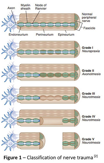
Current non-surgical management involves rest, pain control and resistance training[4]. Though not explored within the literature, neural mobilization may have an important role in patients with persistent symptoms, such as Grade 1 moderate to severe, and more recurrent neuropraxias. Though not assessed in this specific population, there is evidence for neural tissue management being superior to minimal intervention for pain relief and reduction of disability in nerve related chronic musculoskeletal pain.[8] It is biologically plausible that recurrent neuropraxias may respond in a similar way, utilising neural mobilisation (tensioning or sliding) and mobilisation of surrounding structures.

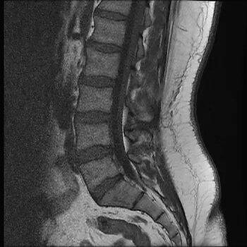
Management of persistent Grade 1 injuries may differ slightly, specifically if the suspected mechanism of injury was through traction rather than compression. The nerve structures may have a heightened sensitivity to tensioning based techniques due to the similar mechanism of injury and may respond better acutely to sliding techniques which limit the strain on the nerve and focus on excursion. Tensioning techniques may be important in the sub-acute phase by loading the patient’s nervous system (i.e. increased strain) in preparation for return to function (i.e. tackling with acute traction on the brachial plexus).

In summary, perhaps we shouldn’t be as dismissive of “stingers”, particularly if they are recurrent for you! If you have any questions or would like to see one of our physios regarding your injury, feel free to contact us on (07) 3102 3337 or book online on our website

Till next time, Praxis what you Preach

Team Praxis

Prevent | Prepare | Perform

REFERENCES:

Menorca, R.M.G., T.S. Fussell, and J.C. Elfar, Nerve physiology: mechanisms of injury and recovery. Hand clinics, 2013. 29(3): p. 317-330.

Tsao B, B.N., Bethoux F, Murray B, Trauma of the Nervous System, Peripheral Nerve Trauma. 6th ed. In: Daroff: Bradley’s Neurology in Clinical Practice. 2012.

Sunderland, S., A classification of peripheral nerve injuries producing loss of function. Brain, 1951. 74(4): p. 491-516.

Ahearn, B.M., H.M. Starr, and J.G. Seiler, Traumatic Brachial Plexopathy in Athletes: Current Concepts for Diagnosis and Management of Stingers. J Am Acad Orthop Surg, 2019.

Feinberg, J.H., Burners and stingers. Phys Med Rehabil Clin N Am, 2000. 11(4): p. 771-84.

Presciutti, S.M., et al., Mean subaxial space available for the cord index as a novel method of measuring cervical spine geometry to predict the chronic stinger syndrome in American football players. J Neurosurg Spine, 2009. 11(3): p. 264-71.

Aldridge, J.W., et al., Nerve entrapment in athletes. Clin Sports Med, 2001. 20(1): p. 95-122.

Su, Y. and E.C. Lim, Does Evidence Support the Use of Neural Tissue Management to Reduce Pain and Disability in Nerve-related Chronic Musculoskeletal Pain?: A Systematic Review With Meta-Analysis. Clin J Pain, 2016. 32(11): p. 991-1004.

FACT OR FICTION FRIDAY || Knee Pain and Scans

FACT OR FICTION FRIDAY || Knee Pain and Scans

Answer: FICTION 🙊

“Osteoarthritis” is a common term that gets used by our patients as an explanation of their knee pain. But is this always the case 🤔?

🔍In a recent systematic review estimates of osteoarthritis feature prevalence on MRI among asymptomatic uninjured knees were up to 14% in adults < 40 years, and up to 43% in adults > 40 years!

Whilst features on MRI imaging such as cartilage defects, meniscal tears and osteophyte lesions can potentially play a role if you have pain, this should always be interpreted in the context of your clinical presentation by a health care professional as these changes can be normal in an asymptomatic population – just like grey hair as we age 👴👵!

If you have knee pain and have resigned yourself to a ‘life sentence’, come and have a chat to one of our physios to ensure you aren’t robbing yourself of a full functioning future 🕺🏃🏌️🏄🏋️🏊🚴🏂🎾! Call 07 3102 3337 or book online 

#kneeoa #praxisphysio #factorfictionfriday #preventprepareperform #kneepain #kneeosteoarthritis #mri #fullfunctionfuture

Reference:

Culvenor AG, Øiestad BE, Hart HF, et al Prevalence of knee osteoarthritis features on magnetic resonance imaging in asymptomatic uninjured adults: a systematic review and meta-analysis British Journal of Sports Medicine 2019;53:1268-1278.

Shin Splints: Causes, Treatment & How to Get Back to Running Stronger

Shin Splints: Causes, Treatment & How to Get Back to Running Stronger

Key Takeaways

  • Shin splints (MTSS) are an overuse bone stress injury.

  • Training load errors are the biggest contributor.

  • Hip strength and force control play a major role.

  • Early management prevents stress fractures.

  • Strength + smart loading beats rest alone.

    Mid Potion Achilles Tendinopathy Location

    Shin Splints

    Shin splints, known in the research as medial tibial stress syndrome (MTSS) are one of the most common running injuries we see at Praxis Physiotherapy across our Teneriffe, Buranda and Carseldine clinics.

    If you’re noticing a dull ache along the inside of your shin that worsens with running, skipping or sport, you’re not alone. The good news? With the right plan, shin splints are highly manageable — and preventable.

    Let’s break down what’s actually happening, why it develops, and what you can start doing today.

    What Are Shin Splints (Medial Tibial Stress Syndrome)?

    Shin splints are an overuse bone stress injury affecting the inner (medial) border of your tibia (shin bone).

    What does it feel like?

    • Aching pain along the lower inside shin

    • Sharp pain when running or jumping

    • Tenderness to touch along the bone

    • Morning stiffness or pain when first starting activity

    Unlike a stress fracture (which is more focal and severe), shin splints usually present as a broader area of tenderness along the bone.

    What’s Actually Happening? (The Pathology Explained Simply)

    MTSS is no longer thought to be just an “inflammation” problem.

    Current evidence suggests shin splints develop due to repetitive loading that exceeds the bone’s capacity to adapt. When running loads increase too quickly, the tibia experiences repeated bending stress. If recovery isn’t adequate, this leads to:

    • Bone stress reaction

    • Irritation of the periosteum (bone lining)

    • Localised pain along the medial tibia

    If ignored, MTSS can progress to a tibial stress fracture, which requires significantly longer time away from running.

    That’s why early management matters.

    Why Do Shin Splints Develop?

    A large systematic review by Winkelmann et al. (2016) identified over 100 potential risk factors for MTSS, with several consistently seen in clinical practice.

    Key Risk Factors Supported by Research

    1. Training Load Errors
    Rapid increases in running volume, intensity, or frequency are one of the strongest contributors.
    (Winters et al., 2013; Nielsen et al., 2012)

    2. Higher Body Mass Index (BMI)
    Greater body mass increases tibial loading forces.

    3. Biomechanical Factors

    • Increased navicular drop (foot pronation)

    • Greater plantarflexion range

    • Hip muscle weakness and poor pelvic control

    4. Previous History of MTSS
    Recurrence risk is higher without proper rehabilitation.

    Importantly flat feet alone are rarely the sole cause.

    What we often see clinically is this chain reaction:

    Poor hip control → knee collapses inward → foot over-pronates → increased traction stress on medial tibia.

    The foot is often the victim, not the culprit.

    Why Rest Alone Isn’t Enough

    Yes — rest reduces pain. But rest does not improve:

    • Load tolerance

    • Running capacity

    • Strength deficits

    • Movement control

    This explains why shin splints are common in recreational runners but less frequent in well-structured training programs. Higher-level athletes typically follow progressive loading plans that allow bone and tendon adaptation.

    Research consistently supports graded loading and strengthening as key components of recovery (Moen et al., 2012).

    .

    How to Manage Shin Splints (Early Stage Tips)

    If your symptoms are mild to moderate, here are evidence-informed starting points:

    1️⃣ Relative Rest (Not Complete Rest)

    Reduce running volume by 30–50%.
    Avoid sharp increases in load.
    Swap some runs for cycling or swimming temporarily.

    2️⃣ Strengthen the “Shock Absorbers”

    Focus on:

    • Calf strength (bent and straight knee)

    • Tibialis posterior strengthening

    • Glute medius and hip control exercises

    • Single-leg stability work

    Improving hip strength can reduce tibial loading by improving force control through the limb.

    3️⃣ Manage Running Workload

    Follow the “10% rule” cautiously.
    Allow recovery days between harder sessions.
    Avoid sudden terrain changes (e.g., grass → concrete).

    4️⃣ Consider Footwear

    Ensure shoes are not worn out (>600–800km).
    Orthotics may help some individuals — but only after assessment.

    When Should You See a Physio?

    Seek professional assessment if:

    • Pain persists longer than 2–3 weeks

    • Pain becomes sharp and localised

    • Hopping on one leg is painful

    • Symptoms worsen despite reducing load

    Early intervention reduces the risk of progression to stress fracture.

    How Praxis Approaches Shin Splints

    At Praxis, we don’t treat “shin splints.”
    We treat your specific loading problem.

    Your plan may include:

    ✔ Comprehensive running and strength assessment
    ✔ Individualised load management plan
    ✔ Targeted strength and control program
    ✔ Manual therapy where appropriate
    ✔ Gradual return-to-run progression
    ✔ Prevention strategy for long-term performance

    Because no two runners load the same way, and no two recovery plans should be identical.

    If you’re dealing with shin splints, or want to prevent them from coming back, our team can help.

    Book an in-depth running and lower limb assessment today.

    📍 Clinics in Teneriffe, Buranda & Carseldine
    📞 (07) 3102 3337
    💻 Book online

    💪 Trusted by athletes. Backed by evidence. Here for everyone.

    The Praxis Team.

    PREVENT | PREPARE | PERFORM

    References

    • Winkelmann ZK et al. (2016). Risk factors for medial tibial stress syndrome in active individuals. Journal of Athletic Training, 51(12), 1049–1052.

    • Winters M et al. (2013). Medial tibial stress syndrome: a critical review. Sports Medicine, 43(12), 1315–1333.

    • Moen MH et al. (2012). Treatment of medial tibial stress syndrome: a systematic review. Sports Medicine, 42(11), 965–981.

    • Nielsen RO et al. (2012). Training errors and running-related injuries. International Journal of Sports Physical Therapy, 7(1), 58–75.


    Images:

    The above images are owned by the “Trainer Academy (https://traineracademy.org/) ” and used in this article with thanks.

    References:

    1. Hopper D, Deacon S, Das S, et al. Dynamic soft tissue mobilization increases hamstring flexibility in healthy male subjects. Br J Sports Med. 2004;39:594–598
    2. Weerapong, P., Hume, P.A. & Kolt, G.S. The mechanisms of massage and effects on performance, muscle recovery and injury prevention. Sports Med 2005; 35: 235
    3. Morelli M, Seaborne DE, Sullivan SJ. Changes in h-reflex amplitude during massage of triceps surae in healthy subjects.J Orthop Sports Phys Ther. 1990;12(2):55-9.
    4. Arroyo-Morales M1, Fernández-Lao C, Ariza-García A, Toro-Velasco C, Winters M, Díaz-Rodríguez L, Cantarero-Villanueva I, Huijbregts P, Fernández-De-las-Peñas C. Psychophysiological effects of preperformance massage before isokinetic exercise. J Strength Cond Res. 2011 Feb;25(2):481-8.

    https://www.massagemyotherapy.com.au/Home