Praxis Represents

Praxis Represents

We pride ourselves on using our skills to help athletes, weekend warriors and every day recreational exercises perform at their best. We also revel in the opportunity to tour and represent Praxis at national and international events.

In the first time in our short history, we have 3 physiotherapists out of the clinic providing their skills for Cricket Australia teams and tournaments both at home and abroad.

Stephen is looking after the Australian U19s in Colombo Sri Lanka, as recently covered in the PWYP blog: A winning Australian Cricket Side. He returns from the subcontinent on the 15th of January.

Zac, is busy down in Shepparton Victoria as a central physiotherapist for the Australia Country Cricket Championships. This competition will provide country cricketers an opportunity to play in a national carnival that will see six teams from across Australia and the East Asia Pacific (EAP) region battling across T20, OneDay and Two-Day cricket. Country cricket continues to be a tremendous nursery for talented young players including national representatives such as premier fast bowlers Andy Bichel and Shaun Tait, showing that this tournament is a viable part of the high performance pathway.

Cameron is about to embark upon two weeks in Ballarat and Bendigo as a central physiotherapist at the U18 Female National Championships. Underage National Championships are an integral part of the Australian Cricket Pathway. These Championships offer important development opportunities for our emerging stars and provide a stepping stone to further national representation. Events such as these have been the major contributor of talent to the Australian Women’s Team since its establishment helping such players as Meg Lanning and Ellyse Perry become the best in the world.

Congratulations to our three physiotherapists and we look forward to seeing them back in clinic soon!

Till next time,

Prevent. Prepare. Perform

Team Praxis
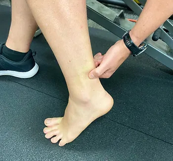
Mid Potion Achilles Tendinopathy Location

Causes and Risks

Achilles tendinopathy typically results from a combination of intrinsic and extrinsic factors. Intrinsic factors include age, reduced flexibility, reduced calf strength / endurance and poor lower limb biomechanics. Extrinsic factors encompass inappropriate footwear, training errors (such as a spike or change in workload), and inadequate warm-up or cool-down routines. Additionally, individuals with systemic conditions like diabetes or rheumatoid arthritis may be more prone to developing Achilles tendinopathy. Understanding these factors is crucial for tailoring treatment plans to address the root causes and minimize the risk of recurrence. But in the most reductionist of terms, Achilles tendinopathy develops due in large part due to a mismatch between loading and the capacity of the tissue.

Diagnosis and Assessment

Accurate diagnosis of Achilles tendinopathy relies on a thorough clinical examination and patient history. Physiotherapists employ various assessment techniques, such as palpation, functional tests, and imaging modalities like ultrasound or MRI, to evaluate the severity and extent of the condition. A self administered questionnaire (VISA-A) can help evaluate symptoms and their effect on physical activity and in turn, the clinical severity. This comprehensive assessment helps determine the appropriate treatment approach, including targeted exercise programs, manual therapy, and other interventions.

Treatment Strategies

Physiotherapy plays a pivotal role in the management of Achilles tendinopathy. Treatment strategies focus on reducing pain, promoting healing, and improving function. These will include calf strengthening exercises, stretching routines and activity modification as frontline options. Moreover, physiotherapists can guide patients in proper footwear selection, gait retraining, and implementing preventive measures to minimize the risk of reinjury.

Rehabilitation and Prevention

Rehabilitation programs are essential for individuals recovering from Achilles tendinopathy. Gradual progression of exercise intensity, functional training, and sport-specific drills enable patients to regain strength, flexibility, and proprioception while minimizing the risk of relapse. Educating patients on proper warm-up and cool-down routines, appropriate footwear selection, and regular monitoring of training loads can significantly contribute to preventing Achilles tendinopathy in the future. One of the common errors patients make is making rehabilitation too easy, or returning to sport too quickly. Again, physiotherapy play a pivotal role in ensuring you undertake a graduated return to loading as the application of mechanical stress to the Achilles tendon promotes tendon healing and remodeling.

Conclusion

Achilles tendinopathy requires a comprehensive approach for effective management. As physiotherapists, our knowledge and expertise are invaluable in helping you overcome this condition and return to their active lifestyles. To discuss your Achilles issues with us to get you back to what you love doing, book online with Praxis today.

Until next time, Praxis What Your Preach.

Team Praxis

A Winning Australian Cricket Side

Apologies for the obviously topical and sensationalist headline, but it is worth mentioning that our principal physio Stephen, is abroad at the moment looking after the Aussie U19 Cricket side in Colombo. The side which was name in mid December, arrived in Colombo on the 30th of December for a two week tournament against their Sri Lankan peers.

Four contracted players including Victoria and Melbourne Renegades duo Will Sutherland and Zak Evans, Tasmania’s Jarrod Freeman and New South Wales’ keeper Baxter Holt were all named in a 14-player squad for the four-match tour of the island nation.

More on the Cricket Australia Announcement here

The Head Coach and former Australian fast bowler Ryan Harris addressed the media on the eve of the series.

The Aussies drew first blood for the series with a composed and convincing win, securing the victory by chasing down Sri Lanka’s 262 with 7 wickets and 2 overs to spare. Sam Fanning was the back bone early with a sensationally well made 108 (94). He was aptly supported by a swashbuckling Ollie Davies who guided the visitors to the finish line with 50*(44) which included 4 massive sixes easily clearing the straight boundaries.

The bowlers toiled hard in typically hot and humid conditions on what appeared to be a batting friendly wicket. Zac Evans and Josh Kann both picked up two wickets whilst the spin of Jarred Freeman and Tanveer Sangha managed to snare 1 a piece.

The remaining two one-day matches will be played at P. Sara Stadium on 5th and 7th January, followed by the three-day match from 10th to 12th January at SSC Grounds.

Live scoring is available via the ESPN cricinfo website.

Good luck to the lads and Stephen for the remainder of the series. Fingers crossed our senior test team can get the job done in Sydney!

Until next time, Praxis what you preach: Prevent. Prepare. Perform

Team Praxis

The Single Leg Squat

The Single Leg Squat

For those of you who have ever read a research article and thought it was a tough read, i’d like to let you in on a little secret. Doing the research is far worse! If only memes were a thing when I started my Masters of Applied Science thesis, I’d have changed my background to remind myself that conducting research was even drier than every dish an apprentice has cooked for Gordon Ramsey.

My mentor at the time, who was the manager of the sports science and sports medicine devision of Cricket Australia and later the head of the AIS human movements department, Dr Marc Portus, enlightened me with something quite profound. He said, “There are two outcomes from a thesis. Either you live it for the rest of you academic days or it sits on a bookshelf for years collecting dust.” Given I completed my thesis and went straight onto my graduate entry physiotherapy masters, it is fair to say i’m in the later camp. Quite a few years have passed now however, so I’d thought i’d dust off the cover and summarise my thesis for all of you playing at home!

So not to degrade my self entirely, I thought I found some pretty good stuff that has affected the way I assess and treat today. I haven’t shared it all today as it was more than 140 pages long and ‘ain’t nobody got time for that’. More importantly though, my thesis reminded me of the passion I have for lower limb biomechanics and that physiotherapy (as opposed to research) was always meant to be on the cards for me. My thesis, “The 3D Kinematics of the Single Leg Flat and Decline Squats” boiled down to looking at how the ankle position changed the joint angles single leg squat, how hip strength affected the squat as well as few other things that aren’t worth mentioning here.

BACKGROUND:

The single leg squat (SLS) replicates an athletic position commonly assumed in sport such as cutting (powerful change in direction while running made from one leg), jumping and balancing which all require the control of the trunk and pelvis on the weight bearing femur in all three planes of movement [1-5].

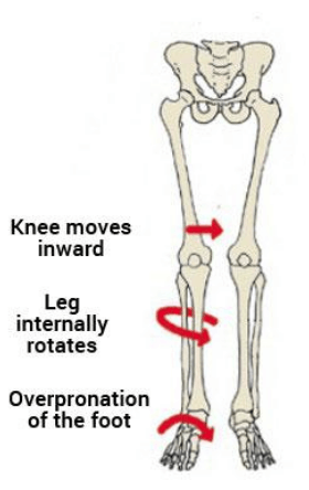
As such, the SLS is commonly used by clinicians as a functional measure of dynamic lumbo-pelvic stability [endif]–[6-8]. Abnormal movement within the SLS tend to be characterised by the commonly described “medial collapse” or “dynamic valgus”. Specifically, there is excessive femoral internal rotation, femoral adduction, knee valgus, tibial internal rotation and foot pronation of the weight-bearing limb with resultant excursion of the contralateral non weight bearing Ilium and excessive lateral flexion of the trunk [endif]–[3, 6-8].

The reason why this tends to be perceived as a big deal is that this position tends to be argued as a lack of lumbopelvic stability and results in increased loading of the knee. Moreover, pelvis weakness tends to be ascribed to the absence of stability ultimately resulting in a position in which many acute and overuse injuries of the lower limb may occur. These ailments include, ACL / MCL ruptures, patellofemoral pain syndrome (PFPS), illiotibial band friction syndrome (ITBFS) and shin splints to name a few. That is why the SLS appears to be a valuable rough screening tool in clinical practice.

MY FINDINGS:

As mentioned, I looked at how a decline board of 20 degrees changed the angles of the lower limb during the squat. I also looked at if any strength measures of the hip related to how someone squatted between conditions. Finally, I looked at if the decline board altered how someone was scored by experienced physiotherapist as a competent or not at the squat

JOINT ANGLES (KINETMATICS:)

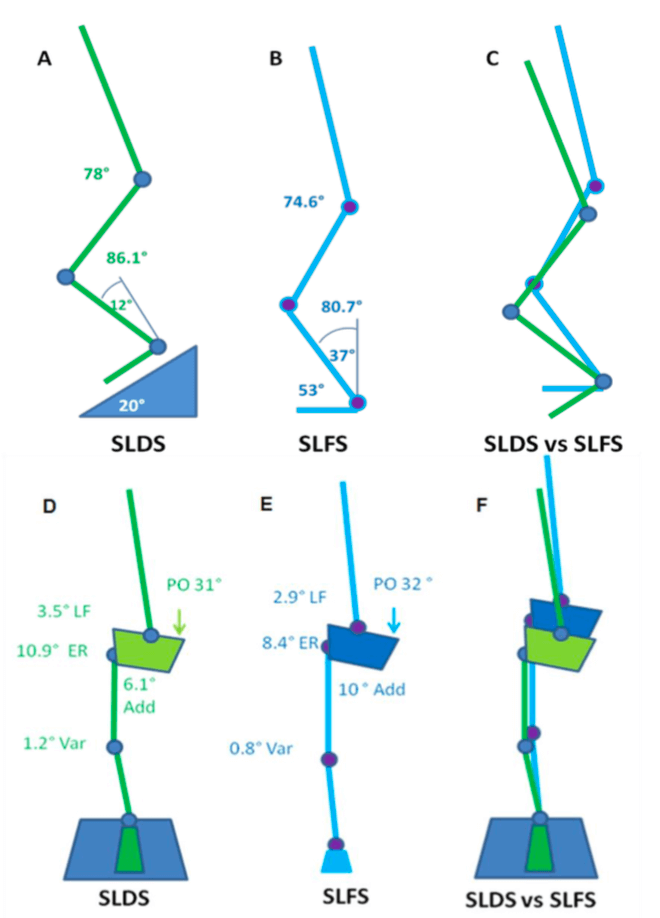
A picture tells a thousand words so in the interests of brevity, the stick squat figure is essentially a summary of two years of work.

So what this means, when someone performs a SLS on a flat surface, relative to a decline surface they tend to have:

  • A more upright torso
  • More rotation of the pelvis toward the weigh bearing (WB) limb
  • Reduced flexion but more adduction and internal rotation of the thigh on the WB hip (pelvic close to femur)
  • Less flexion of the knee but the same position relative to the foot as you look from the front (known as frontal plane knee excursion) at the bottom of range
  • Reduced internal rotation of the shin
  • Reduced ankle flexion

Essentially, in a flat squat you tend to ‘corkscrew’ your pelvis and adopt the medial collapse position much more easily than in the decline squat position. This may because of ankle range of motion issues as well as the ability to adequately recruit pelvic musculature. Yep – two years to get that!

STRENGTH AND MOVEMENT:

My results demonstrated a tendency for the pelvis to remain increasingly level with greater hip abduction strength. However, the relationship between strength and the pelvis was observed in the decline condition but not the flat condition. This may be due to hip abduction was shown to be significantly less (more neutral) in the SLDS which seemingly promoted greater muscle activation and subsequent control of pelvis. The self selection of squat depth may have also been a critical factor in finding as those with weak hips may have squatted deep to adopt maladaptive positions. Previous research has indicated that the hip abductors and external rotators play an important role in lower extremity alignment as they assist in the maintenance of a level pelvis [9] and are capable in balancing a number of biomechanical forces in the body [10].

Interestingly, there were no significant relationships observed between hip abduction strength and knee valgus (knee falling in) for both squatting conditions. There was however a trend between hip abduction strength and knee valgus which supported previous research. It is keeping with the assumption that increased knee valgus might also be associated with reduced hip abduction and external rotation strength [11].

SUMMARY:

  • To maximise athletic function, particularly in sports such as soccer, netball and AFL, stability through the pelvis and hips, proximal lower limb, spine and abdominal structures is required [12].
  • The importance of pelvis stabilisation for lower extremity injury prevention [13] particularly the knee [14-17] has been well documented in the literature.
  • Adequate lumbopelvic-femur strength and muscle function may conceivably reduce exposure to other intrinsic risk factors such as inefficient force attenuation, unstable movement patterns and lower limb malalignments during activity [18, 19].
  • Ankle flexibility may also be a factor in lower limb physical resilience and injury prevention.
  • Support for the previous statements has been demonstrated in the relationships between hip strength measures and kinematics within selected results of my study.

There you have it. Two years of my life summarised to a few paragraphs. From a personal perspective, I took away from my research experience to be always questioning why we do things and see if there is someone out there who has answered the questions we seek. Finally, don’t overcook chicken – Ramsay doesn’t like it.

REFERENCES:

  1. Neely, F.G., Intrinsic risk factors for exercise-related lower limb injuries. Journal of Sports Medicine, 1998. 26(4): p. 253-263.
  2. Parkkari, J., U.M. Kujala, and K. Pekka, Is it possible to prevent sports injuries? Review of controlled clinical trials and recommendations for future work. Sports Medicine, 2001. 31(14): p. 985-995.
  3. Lysens, R.J., et al., The accident -prone and overuse-prone profiles of the young athlete. The American Journal of Sports Medicine, 1989. 17(5): p. 612-619.
  4. Egger, G., Sports injuries in Australia: causes, costs and prevention. A report to the national better health program., ed. C.f.H.P.a. Research. 1990, Sydney.
  5. Orchard, J.W. and C.F. Finch, Australia needs to follow New Zealand’s lead on sports injuries. The Medical Journal of Australia, 2002. 177: p. 38-39.
  6. Wu, G. and P.R. Cavanagh, ISB recommendations for standardization in the reporting of kinematic data. Journal of Biomechanics, 1995. 28: p. 1257- 1261.
  7. Siegal, P., R. Brackbill, and G. Heath, The epidemiology of walking exercise: implications for promoting activity among sedentary groups. American Journal of Public Health, 1995. 85(5): p. 706-710.
  8. Nicholl, J.P., P. Coleman, and B.T. Williams, The epidemiology of sports and exercise related injury in the United Kingdom. British Journal of Sports Medicine, 1995. 29(4): p. 232-238.
  9. Burnet, E.N. and P.E. Pidcoe, Isometric gluteus medius muscle torque and frontal plane pelvic motion during running. Journal of Sports Science and Medicine, 2009. 8: p. 284-288
  10. Niemuth, P., et al., Hip muscle weakness and overuse injuries in recreational runners. Clinical Journal of Sports Medicine, 2005. 15(1): p. 14-21.
  11. Hollman, J.H., et al., Relationships between knee valgus, hip-muscle strength, and hip-muscle recruitment during a single-limb step down. Journal of Sport Rehabilitation, 2009. 18: p. 104-117.
  12. Kibler, W.B., J. Press, and A. Sciascia, The role of core stability in the athletic function Journal of Sports Medicine, 2006. 36(3): p. 189-198.
  13. Leetun, D.T., et al., Core stability measures as risk factors for lower extremity injury in athletes. Medicine & Science in Sports & Exercise, 2004. 36(6): p. 926-934.
  14. Cichanowski, H., et al., Hip strength in collegiate female athletes with patellofemoral pain. Medicine & Science in Sport & Exercise, 2007. 39(8): p. 1227-1232.
  15. Ireland, M.L., et al., Hip strength measures in female with and without patellofemoral pain. Journal of Orthopaedic & Sports Physical Therapy, 2003. 33(11): p. 671-676.
  16. Nicholas, J.A., A.M. Strizak, and G. Veras, A study of thigh muscle weakness in different pathological states of the lower extremity. American Journal of Sports Medicine, 1976. 4: p. 241-248.
  17. Prins, M.R. and P.V.D. Wurff, Females with patellofemoral pain syndrome have weak hip muscles: a systematic review. Australian Journal of Physiotherapy, 2009. 55: p. 9-15.
  18. Willson, J.D., M.L. Ireland, and I. Davis, Core strength and lower extremity alignment during single leg squats. Medicine & Science in Sports & Exercise, 2006. 38(5): p. 945-952.
  19. Lee, D., The pelvic girdle: An approach to the examination and treatment of the lumbopelvic-hip region. 3rd ed. 2004, Edinburugh: Churchill Livingston.
Is running bad for your knees?

Is running bad for your knees?

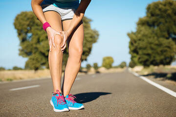
Running. Probably one of the most maligned exercises when it comes to knees and overuse injuries. The thought that running ‘wears’ out your knees and causes osteoarthritis (a chronic disease often associated with joint pain and stiffness, reduced mobility and reduced quality of life) is one of the most common comments I hear as a physiotherapist – typically by non-runners. But do we have it right? Is running actually bad for your knees?

I recently attended the University of Queensland Sports Masters presentation day. The keynote speaker was a Dr Jean-Francois Esculier, a Postdoctoral Fellow at the University of British Columbia on the topics of running and knee osteoarthritis. Originally trained as a physiotherapist, Dr Esculier gave us an excellent overview of his latest research his take on whether or not running is detrimental to knee health.

First, there was an acknowledgement that echoed the sentiments in the opening paragraph. A study in which Dr Esculier undertook attempted to ascertain the perception about running and the knee joint health among the public and health care professionals. The results suggested that many non-runners perceived running as detrimental to knee health. Understandably, with no clear guidelines, health care professionals displayed high rates of uncertainty regarding running as a risk factor to develop knee osteoarthritis (KOA), and about the appropriateness of running with pre-existing KOA [1].

Mid Potion Achilles Tendinopathy Location

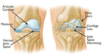
Osteoarthritis often results in cartilage loss, in bone rubbing on bone, which can cause inflammation, pain, stiffness, reduced mobility and reduced quality of life [7].

The paucity of clear training parameters for runners also has a knock on effect with a staggering 75% of runners report being injured whilst running each year with the knee being the most common region of complaint [2]. From my experience as a clinician, the factor that is most often associated with an injury are training errors. Too much too quick. Boom bust. No physical preparation. No listening to your body or allowing adequate recovery time. No periodisation or plan – just run and run.

The remainder of the talk that covered many interesting relatable topics (that will likely be areas for future blogs) but the information that most interested me and should answer the question as to whether running is bad for your knees was the following:

Cartilage change with running:

With the improvements in MRI scanning, more papers are looking at the cartilage volume of knees immediately after a long distance run. According to current evidence [3], cartilage may exhibit short-term decreases in thickness, volume and cartilage water flow (T2 relaxation time) secondary to temporary loss of fluid following repeated compressions associated with running. However, cartilage size tends to return to baseline within hours suggesting that cartilage may well tolerate mechanical loading sustained during running and adapt to repeated exposure.

The response of cartilage to longitudinal load is exactly what Van Ginckel et al [4] investigated. After providing a 10 week “Start To Run” program to novice runners, the reserachers looked at the glycosaminoglycan (GAG) content before and after the running intervention and compared to sedentary controls, who did no running. For those of you (like me) who had no idea what glycosaminoglycan / GAG content is, it is essentially a surrogate marker for cartilage quality (specifically, GAG is an important structural matrix compound in regulating the cartilage tissue’s endosmotic swelling pressure and thus, the tissue’s compressive strength).

The results suggested that a gradually built up running scheme appears to positively effect GAG content, and thus cartilage quality. In fact, running appears to be a chondroprotective effect on the knee when compared to a sedentary lifestyle in a female asymptomatic subjects. The author’s went onto say that running schemes like this might be considered a valuable tool in osteoarthritis prevention strategies [4].

Osteoarthritis (OA) rates in competitive vs recreational vs non-runners:

The body’s ability to adapt to considered and appropriate load is likely explanation as to why when we look at the rates of lower limb osteoarthritis (OA) across the population, we find some interesting results. A systematic review [5] of the literature looked at the association of recreational and competitive running with hip and knee OA. The overall prevalence of hip and knee OA was 13.3% in competitive runners, 3.5% in recreational runners, and 10.2% in controls. Exposure to running of less than 15 years was associated with a lower association with hip and/or knee OA compared with non-runners.

Recreational runners had a lower occurrence of OA compared with competitive runners and controls. These results indicated that a more sedentary lifestyle or long exposure to high-volume and/or high-intensity running are both associated with hip and/or knee OA. However, it was not possible to determine whether these associations were causative or confounded by other risk factors, such as previous injury [5].

SUMMARY

Running appears not to cause osteoarthritis in your knees unless you are a competitive long distance runner. Even then, you are only slightly above the average for non-runners but enjoy the myriad of other benefits that exercise brings. Further, increased mileage in recreational runners appears to be actually protective for your knees and reduces your risk of needing a knee replacement [6]. Caution however must be taken to monitor detailed training parameters such as frequency, speed and distance, so that an optimal dosage for knee joint health tailored to the individual patients with knee osteoarthritis.

So the next time someone tells you that you shouldn’t be running because you’ll get OA, or if your health expert recommends to stop all activity because you have been diagnosed with mild / moderate osteoarthritis of the knee, we can help! As always, we at Praxis are more than happy to help you navigate your way back to performing – whatever that may look like! Give us a call (07) 3102 3337 or book online www.praxisphysio.com.au today

Until next time, Praxis What You Preach

There is no need to accept knee pain as ‘normal’. Call us now on (07) 3102 3337 or book online to have one of our physios develop a plan to reduce your pain and restore your function!

To read more about how running can help your knees (that’s right – running!) check out our related posts on running written by our published principal physio, Stephen.

Team Praxis,

PREVENT | PREPARE | PERFORM

Why lifting is your missing endurance link: A guide for long distance runners (Part 1)

Why lifting is your missing endurance link: A guide for long distance runners (Part 1)

You have the shoes, the GPS watch, training schedule and alarm set for 5am. You are dedicated and that race is right around the corner. Whether it is your first 5km or your 50th marathon, the thrill of crossing the finish line drives us all. Whilst you may know your average km split time like the back of your hand, do you know how strong your lunges or deadlifts are? If you haven’t stepped foot in a gym recently, then research suggests you could be missing out on a host of positive effects on your running.There has been a whole host of research in this area so deciphering the literature can be a difficult task. Thankfully, a recent paper by Blagrove et al [1] has done much of the hard work for us. The paper entitled Effects of Strength Training on the Physiological Determinants of Middle- and Long-Distance Running Performance: A Systematic Review aimed to provide a comprehensive critical commentary on the current literature that has examined the effects of strength training modalities on the physiological determinants and performance of middle and long-distance runners. They also offered recommendations for best practice which you can read about in the Part 2 blog post.
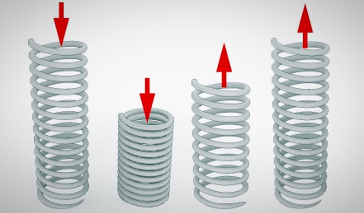
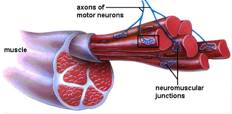
Running is a surprisingly complex task and as such there are many factors that affect performance. Physiological, biomechanical, psychological, environmental, and tactical factors all inter play to result in determining the average runner from the elite. With respect to physiological markers of performance, maximal oxygen uptake (known as VO2max), running economy, and the sustainable percentage of VO2max go a long way to determining performance [2]. In fact, these three elements can predict performance with up to 95% accuracy in well trained runners.The difference between VO2max in the elite running population however is surprisingly marginal. On the contrary, running efficiency displays a high degree of inter-individual variability and thus a potential area to better discriminate between runners and their respective performance [3]. Defined as the oxygen or energy cost of sustaining a given sub-maximal running velocity, running efficiency is underpinned by a variety of anthropometric, physiological, biomechanical, and neuromuscular factors [4]. More specifically to the purpose of this article, force generation and stretch–shortening cycles are the neuromuscular factors that are the most relevant.Whilst force production of a muscle is a straight forward concept, the stretch shortening cycles may not be. Stretch shortening cycles describe the pre-stretch and recoil action of a muscle and tendon unit that occurs in a dynamic action just as jumping. Think of the stretch shortening cycle like a spring whereby energy is stored and released within the spring, or in real terms, the musculo-tendinous unit. To produce higher forces, the more motor units (muscle) are required [5]. There is a strong correlation between the cross-sectional area of a muscle and its ability to produced force. Several other factors are involved, but for the most part, a larger muscle will produce more force than a smaller muscle. However, force production becomes more difficult when activities are dynamic. This is because there is a reduction in force produced per motor unit due to the faster shortening velocity involved in the stretch shortening cycle [5].In general, strength training activities can positively affect both muscle force as well as improve the stretch-shortening cycle through several different adaptations including muscular and neural changes [6, 7]. Hypertrophy is the term to describe an increase in muscle size. It is the cyclical process whereby muscle cells are exposed to repeated bouts of exercise causing micro damage to the muscle cells. Micro damage causes an inflammatory response and it is the pain you feel for the next 48hrs after a bout of exercise (also called delayed onset muscle soreness or DOMS for short). It is also the stimulus for the body to mitigate future damage by repairing the damaged tissue and adding more muscle cells. This is what is commonly known as the super compensation cycle. Hypertrophy is aided by rest, dietary protein, certain hormones (e.g testosterone) and has a very strong genetic component as well [7].
Neural adaptation tends to be one of the earliest changes and accounts for most of the strength increases observed in the initial stages of all strength training [8]. Those who are exposed to repeated bouts of resistance training generate significant strength gains with minimal hypertrophy early in the process. The body achieves this via synchronous activation (the ability to recruit more muscle cells in a simultaneous fashion) and reduction in neural inhibition (a natural response of the central nervous system to feedback signals arising from the muscle) [9]. Inhibition allows muscle to avoid overworking and potentially damaging itself due to unaccustomed load. This response is rapid as it utilises the nerve and muscle cells already present. These adaptations are in direct contrast to the untrained muscle in which atrophy (muscle wastage) and reduced neural drive are typical.What this all boils down to is that following a period of strength training there is an increase in absolute motor unit recruitment resulting in a lower relative intensity of that muscle unit to deliver the same outcome as previous. If the bouts are habitual and frequent enough, muscle cells hypertrophy and become larger, increasing their ability to generate force. As a result, the trained muscle will be able to recruit a higher threshold of larger motor units. Combine all of this with an enhanced stretch shortening cycle and you have some excellent adaptations to improve running efficiency.

With respect to the dosage, the Blagrove paper suggested, a strength training intervention, lasting 6–20 weeks, added to the training program of a distance runner appears to enhance running efficiency by 2–8%. In real terms, an improvement in running efficiency of this magnitude should theoretically allow a runner to operate at a lower relative intensity and thus improve training and/or race performance. Improvements were observed in moderately-trained, well-trained and highly-trained participants, suggesting runners of any training status can benefit from strength training. For the particulars of the dosage, exercise selection and periodisation, check out Part 2 blog post.

Until next time, continue to Praxis What You Preach…

Prevent. Prepare. Perform.

References:

  1. RC. Blagrove, G Howatson, PR. Hayes. Effects of Strength Training on the Physiological Determinants of Middle- and Long-Distance Running Performance: A Systematic Review, Sports Med. 2018; 48(5):1117-1149
  2. McLaughlin JE, Howley ET, Bassett DR Jr, et al. Test of the classic model for predicting endurance running performance. Med Sci Sports Exerc. 2010;42(5):991–7
  3. Morgan DW, Craib M. Physiological aspects of running economy. Med Sci Sports Exerc. 1992;24(4):456–61.
  4. Saunders PU, Pyne DB, Telford RD, Hawley JA. Factors affecting running economy in trained distance runners. Sports Med. 2004;34(7):465–85.
  5. Barnes KR, Kilding AE. Running economy: measurement, norms, and determining factors. Sports Med. 2015;1(1):8–15
  6. Denadai BS, de Aguiar RA, de Lima LC, et al. Explosive training and heavy weight training are effective for improving running economy in endurance athletes: a systematic review and meta-analysis. Sports Med. 2017;47(3):545–54
  7. Schoenfeld BJ, Ogborn D, Krieger JW. Effects of resistance training frequency on measures of muscle hypertrophy: a systematic review and meta-analysis. Sports Med. 2016;46(11):1689–97
  8. Aagaard P , Simonsen EB , Magnusson SP , Andersen JL , Dyhre-Poulsen P. .Enhanced motoneuron activation as effect of heavy-resistance strength training in man.Med Sci Sports Exerc 29: S23-1997.
  9. Aagaard, P., E. B. Simonsen, J. L. Andersen, S. P. Magnusson, J. Halkjær-Kristensen, and P. DyhrePoulsen. Neural inhibition during maximal eccentric and concentric quadriceps contraction: effects of resistance training. J Appl Physiol 89: 2249–2257, 2000