Peri-Menopause and injury – your guide to the most common issues

Peri-Menopause and injury – your guide to the most common issues

  • Menopause alters hormones and results in physical changes
  • These changes typically result in increased injury risk or activity reduction
  • The most common injuries affect structures such as the plantar fascia, tendons of the hip and shoulder
  • Appropriate exercise and judicious hands on therapy should be cornerstones of management
Betty Friedan said it best when she said “Aging is not lost youth but a new stage of opportunity and strength.” If only our bodies would play the game and come along for the ride as Betty wishfully thought. Age related transitions such as menopause often results in physiological changes (as outlined in other blogs) is an increase in weight, a loss of muscle and bone mass and a drop in physical activity. Not something anyone would wait in line for. A spiral of further activity loss, deconditioning and ultimately, injury can easily occur as a result. As a physiotherapist who looks after everyone from elite athletes, to weekend warriors to desk jockeys, I see the frustration injuries cause my patients (and myself!). So, in the interests of helping you enter the chapter of “opportunity and strength”, these are the most common injuries I see on a daily basis in perimenopausal women and what to do about them.

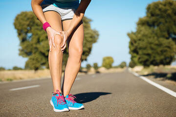
Heel pain – Plantar fasciopathy (the injury formerly known as plantar fasciitis)

Heel pain can be a number of things. The heel pain that I tend to see the most of is the one that feels like you are walking on glass the first thing in the morning or after you have been sitting down for a while. It makes you walk like your Grandma used to for those first few steps and the thought of doing a long walk, or heaven forbid wearing heels, gives you the sweats.

This pain is usually characterized by a condition called plantar fasciopathy. The plantar fascia is designed to help to absorb, store and transfer force during walking, running and jumping activities. Collagen (the main protein based building blocks of the body) is a non contractile tissue that sits on the underside of your foot. The attachment point for this tissue, is you guessed it, on the bottom of your heel where that “burning glass” feeling .

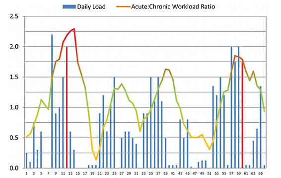
Plantar fasciopathy loves the status quo. It likes loading the same way, at the same intensity, at the same volume day in day out. Pain and dysfunction is brought about typically in a sudden increase in dynamic loading, whether that is walking, running or jumping. This often happens with women hell bent on a health kick to lose those extra kilos. With the shoelaces tied, lycra on and the world your oyster, some women start at where they remember they left their exercise behind, often several years (and children) ago. The resultant spike in load, particularly dynamic loading coupled with musculoskeletal detraining, is what kicks off the pain as the plantar fascia gets overloaded more easily and grizzles about it.

Key tip – Start slow with walks – not running – if it has been a while since you last exercised. Progress to jogging with some walk intervals in there (e.g 30s jog, 90s walk). Do calf raises (eg. 3 sets to a feeling of semi-fatigue) to get more calf strength as this can help deload the plantar fascia.

Outside / Lateral hip pain (Gluteus medius tendinopathy)

The muscles on the outside of your hips are designed to help keep your pelvis level when you are walking or running. This area is a particular passion of mine given my masters thesis was based on the single leg squat. Similar to the plantar fasciopathy, gluteal tendinopathies tend to be an injury that I see when people go too hard, too quickly. This results in outside hip pain that hurts rising from a chair, walking up stairs or even lying on that side at night.

The human instinct is to rest, but tendinopathies don’t work that way, in fact it is the opposite. The only way to adequately address issues such as gluteal tendinopathy is getting on top of your workload. Ensure you are graduating whatever activity you are re-engaging in appropriately, and you have the strength to do so. I typically use a hand held dynamometer in my clinic to assess someone’s strength to give me accurate numbers to work with. However, a rough guide is that if you can do a side plank for 30 seconds, you need some more hip strength.

Key tip – Workload management and pain management are paramount. If in pain, reduce loading and focus on strength. Lying hip lifts (e.g. 10 x 10 sec holds) are good when pain is acute. If not sore (just weak), side plank with top leg lifts are a good option to start. 3 x semi-fatigue each side. We employ reformer pilates often in this space but gym based activities are also appropriate.

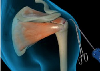
Shoulder Pain – “Rotator Cuff” tears / tendinopathy

Do you have a ‘good’ and ‘bad’ shoulder? Have you been putting up with that grumbly shoulder for weeks, months or even years? Shoulder pain can put a real dampener on activities where repetitive overhead dynamic loading is common (e.g weights, tennis, golf or boxing).

The “rotator cuff” is a group of four muscles that help provide stability and control of the shoulder joint through range, particularly overhead. Rotator cuff tears are common in individuals over the age of 40 with linear increase in incidence as we get older. The most common reason for rotator cuff tears are due to overactivity of the shoulder joint coupled with deconditioning of the shoulder complex. Pain with movement and function is one of the biggest symptoms of a rotator cuff pathology.

Key Tip – Look at how much overhead activity you are doing and adequately prepare for it. This is best achieved in my experience with something like a banded Y Press hold (10x10s holds daily for a month) then progressing to more dynamic activities.

All in all, changes associated with menopause can leave you open to more injuries. However, if you can appropriately navigate, and most importantly graduate, your return to physical activity, then your body will certainly pay you dividends down the line. The evidence is overwhelming for the positive influences physical has on the human body. So do yourself (and your future self) a favour and ensure you aren’t retreating from your physical activity goals due to injury. It’s not what Betty would want.

If you have some of the above aches and pains, feel free to reach out or book in with one of our expert therapists.

Until next time, Praxis what you Preach.

Stephen Timms

All information is general in nature and it is always best to see your local physiotherapist, who uses exercise as the cornerstone of your rehabilitation.

Knee Pain

Knee Pain

Do you have a good knee and a bad knee? Do your knees snap, crackle and pop? Do you ever wish you could walk for longer without pain or not be worried about how your knees will feel in the morning if you were living your life to the fullest? Knee pain is a very common condition that affects people of all ages. Typically knee pain is the result of an injury on the sporting field when we are younger but tend to be more degenerative in nature as we age.
Mid Potion Achilles Tendinopathy Location

Different knee injuries tend to have differing symptoms. Common symptoms include:

  • Aching, sharp, stabbing and/or catching pains
  • Large amounts of swelling or sometimes pockets of swelling
  • Warm to touch
  • Feelings of grating, grinding or even giving way
  • Pops and crunching noises
  • Unable to full bend or straighten the knee

The structures of the knee that are often implicated in knee pain are the patellar or quadriceps tendons, cartilage, meniscus, bursas, and even major ligaments such as the anterior cruciate ligament, otherwise known as the ACL.

Common injuries to the knee can be:

  • Osteoarthritis
  • Tendinopathy / Tendonitis
  • Bursitis
  • Knee cap pain (patellofemoral pain)
  • Meniscal tear (degenerative and acute tears)
  • Dislocated knee cap
  • Iliotibial band friction syndrome

Regardless of your symptoms and presentation, our highly trained Praxis physiotherapists have expertise in this area and will help identify the problem and work with you so that you will feel empowered to fix the problem. At Praxis Physio this is our point of difference, we promise to take the time to fix you using a range of modalities including advice, hands on manual therapies and of course strength and conditioning programming. In addition, we work in close collaboration with leading knee surgeons if this course of action is required.


There is no need to accept knee pain as ‘normal’. Call us now on (07) 3102 3337 or book online to have one of our physios develop a plan to reduce your pain and restore your function!

To read more about how running can help your knees (that’s right – running!) check out our related posts on running written by our published principal physio, Stephen.

Team Praxis,

PREVENT | PREPARE | PERFORM

FACT OR FICTION FRIDAY || All rotator cuffs tears need surgery.

FACT OR FICTION FRIDAY || All rotator cuffs tears need surgery.

Answer: FICTION. The devil is in the detail!

The rotator cuff is a group of 4 muscles that aid in providing stability through range for the shoulder joint, particularly overhead. There are multiple risk factors for RC tears, but most are down to overactivity of the shoulder joint decreased conditioning of the shoulder complex, which comes with age. In fact, cuff tears are common in individuals over the age of 40 with linear increase in incidence as we get older.

Pain with movement and function is one of the biggest symptoms of a rotator cuff tear. However, it is important to know that a sizeable portion of RC tears are actually asymptomatic and don’t cause the person any pain or discomfort! A study by Minagawa and Yamamoto in 2013 found that in a screening of 664 village residents, 147 subjects had RC tears on a medical imaging screening. Surprisingly 65% of them had no symptoms at all and didn’t have any shoulder complaints.

Mid Potion Achilles Tendinopathy Location

The two main ways of treating a cuff tear is either through conservative management with your physiotherapist or down the surgical route, which is also then followed by physiotherapy rehabilitation.

SO, back to the original question: “Do I need surgery?”. As always – it is a case by case decision!! There is no definitive evidence for supporting one over the other generally speaking!

However, it is vitally important to note that each option comes with their own pros and cons. Furthermore, it is important to remember that just as every person is different, each case of rotator cuff tear is different. Young vs old, acute vs degenerative RC tear, current and desired future function, pain levels, radiographic findings, previous history of shoulder trauma and the patient’s wishes are only some of the questions that aid in the decision process.

The best way to decide would be to contact us to asses you and discuss both options and what your goals of rehab are so that a tailored plan can be developed WITH you. To read more about RC tears, read our blog here

To get your shoulder back on track, book online or give us a call on (07) 3102 3337.

Team Praxis

PREVENT | PREPARE | PERFORM

FACT OR FICTION FRIDAY || Overuse injuries need rest and are because I’m doing too much

FACT OR FICTION FRIDAY || Overuse injuries need rest and are because I’m doing too much

ANSWER: FICTION (Mostly – stay with us here) 🙊Do you keep getting injured when you get back into your usual training after a period of rest 😤? The first graph shows a 65-day cycle of an Olympic athlete. The red lines indicate when the athlete was injured. Looking at the multi-coloured line, you can see that the injuries both occurred when the acute to chronic workload ratio was at its peak. Essentially that means that the loads that preceded the injury were too high relative to the longer term loading of the tissue 😫. This is typically called an overuse injury!Then along comes Mr/Ms physio and looks at the above cycle and says to the Olympian, “Come on mate, we can do better than this…” 😎Ta da. The second graph shows the yellow section outlining the above 65-day period.
Mid Potion Achilles Tendinopathy Location

As you can see, training following this period is much more frequent and at higher intensities. So do we still assign the original injuries to overuse injuries given the athlete was able to handle much more relatively quickly after?! A better term would be a training load error and something that a sporting physio can help you with 👌. This is a particularly important thing to know given the holidays are just around the corner 🎅!

Get in touch with us if you want to train more with less injuries, or are looking to return to training! 📞(07) 3102 3337 or book online.

Team Praxis

PREVENT | PREPARE | PERFORM

Drew, M. K., & Purdam, C. (2016). Time to bin the term ‘overuse’ injury: is ‘training load error’ a more accurate term? Br J Sports Med, 50(22), 1423. doi:10.1136/bjsports-2015-095543

Stingers AKA Neural traction injuries

Stingers AKA Neural traction injuries

SUMMARY:

  • Stingers are essentially a tractioning of the neural system
  • This can cause pain, movement and sensation changes
  • Chronic traction to the nervous system can have a cumulative effect on nerve function
  • The more damage to the nerve, the more serious the outcome
  • We shouldn’t be as dismissive of “stingers”, particularly if they are recurrent
  • Physiotherapy has a role to play

STINGERS:

Stingers have been the catch cry of many contact sports over the years. Often dismissed as ‘just a stinger’, trauma to the nervous system should probably be taken a little more seriously, as we delve into detail today.

Stingers are most commonly experienced in contact sports whereby the shoulder of a players is forcefully depressed, as experienced with a tackle in NRL or union. A large range of motion over a short period of time can result in a ‘traction’ of the Brachial Plexus (a network of nerves formed from exiting branches of the spinal cord in the neck that transverse to the shoulder and arm). Thus network of nerves sends signals from your spinal cord to your shoulder, arm and hand and thereby providing feeling and movement to these regions.

BACKGROUND:

To understand a stinger injury, a clear understanding of nerves and nerve related injuries is required. A nerve is an enclosed, cable-like bundle of nerve fibres called axons, in the peripheral nervous system. A nerve provides a common pathway for the electrochemical nerve impulses that provide a number of functions, including getting our muscles to move!

Seddon and Sunderland present a five-grade classification scale for nerve related injuries[1-4]. Figure 1 illustrates the physiological changes that occur through each grade of injury. Essentially it outlines the greater amount of disruption to the anatomy of the nerve.

The more damage to the nerve, the more serious the outcome. Table 1 outlines the three different grades of stinger injuries.[4, 5] The most common stinger is a grade 1 injury, which represents a neurapraxia, or nerve stretch injury, without axonal disruption.[4] In an acute setting, this can result in motor and sensory loss/changes, which usually resolve within minutes.[1, 4] Grade 2 and 3 involve a higher degree of nerve injury, usually involving a crush, transection or compression mechanism.[1]

Chronic traction to the nervous system can have a cumulative effect on nerve function. This is termed “chronic stinger syndrome” and represents a distinct entity from acute stingers that may reflect long- standing structural changes of the subaxial spinal canal and chronic irritation/degeneration of the exiting nerve root complex.[4, 6]. This sounds complex but essentially means

Mid Potion Achilles Tendinopathy Location

A Clinical Example from Zac

“During a Gridiron match, a player was injured whilst making a tackle. I reviewed the player on field and he was unable to utilise his right upper limb (full paralysis) from shoulder down to his hand. The player was removed from the field immediately to be further assessed and monitored. A complete neurovascular assessment was performed, assessing motor function/strength, sensation, reflexes, and vascular status, as well was the cervical spine. Motor and sensation changes were the only deficits noted and were reviewed frequently. After roughly five minutes, the player demonstrated full upper limb motor strength and sensation, with nil lingering symptoms. In collaboration with the patient, it was decided he would return to match play immediately. The patient was monitored throughout the game and reported no further symptoms.”

Figure 2 shows a proposed decision tree when managing stinger injuries.[4] This clinical example outline above fits the Grade 1 Mild category as he was able to return to competition with nil lingering symptoms. Despite the lack of symptoms during the game, it is recommended the patient be reviewed again both after the game and weekly for two weeks to ensure a full resolution of symptoms.[4, 7]

The role for neural mobilisation?

Current non-surgical management involves rest, pain control and resistance training[4]. Though not explored within the literature, neural mobilization may have an important role in patients with persistent symptoms, such as Grade 1 moderate to severe, and more recurrent neuropraxias. Though not assessed in this specific population, there is evidence for neural tissue management being superior to minimal intervention for pain relief and reduction of disability in nerve related chronic musculoskeletal pain.[8] It is biologically plausible that recurrent neuropraxias may respond in a similar way, utilising neural mobilisation (tensioning or sliding) and mobilisation of surrounding structures.

Management of persistent Grade 1 injuries may differ slightly, specifically if the suspected mechanism of injury was through traction rather than compression. The nerve structures may have a heightened sensitivity to tensioning based techniques due to the similar mechanism of injury and may respond better acutely to sliding techniques which limit the strain on the nerve and focus on excursion. Tensioning techniques may be important in the sub-acute phase by loading the patient’s nervous system (i.e. increased strain) in preparation for return to function (i.e. tackling with acute traction on the brachial plexus).

In summary, perhaps we shouldn’t be as dismissive of “stingers”, particularly if they are recurrent for you! If you have any questions or would like to see one of our physios regarding your injury, feel free to contact us on (07) 3102 3337 or book online on our website

Till next time, Praxis what you Preach

Team Praxis

Prevent | Prepare | Perform

REFERENCES:

Menorca, R.M.G., T.S. Fussell, and J.C. Elfar, Nerve physiology: mechanisms of injury and recovery. Hand clinics, 2013. 29(3): p. 317-330.

Tsao B, B.N., Bethoux F, Murray B, Trauma of the Nervous System, Peripheral Nerve Trauma. 6th ed. In: Daroff: Bradley’s Neurology in Clinical Practice. 2012.

Sunderland, S., A classification of peripheral nerve injuries producing loss of function. Brain, 1951. 74(4): p. 491-516.

Ahearn, B.M., H.M. Starr, and J.G. Seiler, Traumatic Brachial Plexopathy in Athletes: Current Concepts for Diagnosis and Management of Stingers. J Am Acad Orthop Surg, 2019.

Feinberg, J.H., Burners and stingers. Phys Med Rehabil Clin N Am, 2000. 11(4): p. 771-84.

Presciutti, S.M., et al., Mean subaxial space available for the cord index as a novel method of measuring cervical spine geometry to predict the chronic stinger syndrome in American football players. J Neurosurg Spine, 2009. 11(3): p. 264-71.

Aldridge, J.W., et al., Nerve entrapment in athletes. Clin Sports Med, 2001. 20(1): p. 95-122.

Su, Y. and E.C. Lim, Does Evidence Support the Use of Neural Tissue Management to Reduce Pain and Disability in Nerve-related Chronic Musculoskeletal Pain?: A Systematic Review With Meta-Analysis. Clin J Pain, 2016. 32(11): p. 991-1004.