Unilateral vs Bilateral Training: Part 1 – Your Comparative Guide

Unilateral vs Bilateral Training: Part 1 – Your Comparative Guide

We often get asked what are the benefits of one form of exercise versus another. Sometimes this is as simple as discussing the difference between cardio and weights. Today’s article focuses on the difference between a two types of gym based loading programs Bilateral vs Unilateral training. In Part 2, we delve more into some of what the evidence says regarding these modalities.

In general, strength training plays a pivotal role in athletic development, enhancing performance, preventing injuries, and promoting overall fitness. This is especially true for the injured population. As such, our Praxis physiotherapists will discuss with you how your rehabilitation plan will incorporate strength training.

When it comes to strength training though, two primary approaches are often employed: unilateral and bilateral training. Unilateral training focuses on exercises that isolate a single limb or side of the body, whereas bilateral training involves movements that engage both limbs simultaneously. This blog aims to contrast the benefits and drawbacks of unilateral and bilateral strength training methods, examining their impact on athletic performance and overall physical development.

Unilateral Strength Training

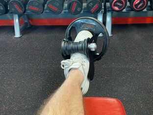
Unilateral strength training involves exercises that emphasize working one limb or side of the body independently. One of the significant advantages of unilateral training is its ability to identify and correct muscle imbalances. By targeting each limb separately, athletes can pinpoint weaknesses, imbalances, or asymmetries, and address them with specific exercises. Unilateral training also enhances proprioception and balance by requiring greater neuromuscular control. It activates stabilizer muscles and enhances coordination, which can lead to improved athletic performance and injury prevention.

Moreover, unilateral training allows for greater range of motion and flexibility development, as each limb can move freely without the restrictions imposed by bilateral movements. This can be particularly beneficial for athletes who need to improve mobility and functional strength in specific joints or muscle groups. Additionally, unilateral exercises offer sport-specific advantages by simulating movements that athletes encounter during competition, such as single-leg jumps in basketball, change of direction football or one-arm strokes in swimming.

However, unilateral training does have limitations. It generally requires more time and effort to complete a full-body workout due to the need to perform exercises separately for each limb. Additionally, unilateral exercises tend to involve lower weight loads, which may limit their potential for developing maximum strength.

Bilateral Strength Training

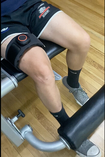
Bilateral strength training, on the other hand, focuses on exercises that engage both limbs simultaneously. One of the primary benefits of bilateral training is the ability to lift heavier weights. This can lead to significant gains in maximal strength and power, making it particularly advantageous for athletes involved in sports that require explosive movements, such as weightlifting or sprinting.

Mid Potion Achilles Tendinopathy Location

Bilateral exercises also promote increased overall muscle mass and hypertrophy due to the higher loading potential. By engaging multiple muscle groups simultaneously, bilateral training can provide a time-efficient method for achieving muscle growth and development. Additionally, the bilateral movements help improve intermuscular coordination, allowing athletes to transfer strength gains more effectively across various activities.

However, bilateral training may not address asymmetries or imbalances as effectively as unilateral training. In some cases, stronger limbs may compensate for weaker ones, perpetuating muscle imbalances and potentially increasing the risk of injury. Moreover, bilateral exercises may not fully translate to specific sport-related movements that often require unilateral actions.

In summary, both unilateral and bilateral strength training methods offer unique benefits and drawbacks for athletes. Unilateral training aids in identifying and correcting muscle imbalances, enhancing proprioception, and improving sport-specific movements. It is a valuable tool for injury prevention and rehabilitation. On the other hand, bilateral training allows athletes to lift heavier weights, develop overall muscle mass, and enhance intermuscular coordination. It is particularly effective for activities that demand explosive power. Ultimately, the choice between unilateral and bilateral training should be determined by an athlete’s specific needs, goals, and the demands of their respective sport. Further, an accurate assessment of any asymmetries that may be present helps to decide where to start. A well-rounded strength training program can incorporate elements of both methods to optimize performance and minimise the risk of injuries.

To read more about the specifics of athletic performance, read our Part 2 Blog.

To help with the genesis of a training program or to chat about your training your goals, book in with one of our knowledgeable Praxis physiotherapists. We are here to help!

Until next time,

Praxis What You Preach

Treatment Strategies

Physiotherapy plays a pivotal role in the management of Achilles tendinopathy. Treatment strategies focus on reducing pain, promoting healing, and improving function. These will include calf strengthening exercises, stretching routines and activity modification as frontline options. Moreover, physiotherapists can guide patients in proper footwear selection, gait retraining, and implementing preventive measures to minimize the risk of reinjury.

Rehabilitation and Prevention

Rehabilitation programs are essential for individuals recovering from Achilles tendinopathy. Gradual progression of exercise intensity, functional training, and sport-specific drills enable patients to regain strength, flexibility, and proprioception while minimizing the risk of relapse. Educating patients on proper warm-up and cool-down routines, appropriate footwear selection, and regular monitoring of training loads can significantly contribute to preventing Achilles tendinopathy in the future. One of the common errors patients make is making rehabilitation too easy, or returning to sport too quickly. Again, physiotherapy play a pivotal role in ensuring you undertake a graduated return to loading as the application of mechanical stress to the Achilles tendon promotes tendon healing and remodeling.

Conclusion

Achilles tendinopathy requires a comprehensive approach for effective management. As physiotherapists, our knowledge and expertise are invaluable in helping you overcome this condition and return to their active lifestyles. To discuss your Achilles issues with us to get you back to what you love doing, book online with Praxis today.

Until next time, Praxis What Your Preach.

Team Praxis

Stress Fractures in Young Cricket Fast bowlers: A persistent challenge

Stress Fractures in Young Cricket Fast bowlers: A persistent challenge

Lumbar stress fractures are one of the most serious and persistent injuries affecting cricket fast bowlers, especially in adolescents. The repetitive, high-load forces of the bowling action place significant strain on the lower back—particularly the L4 and L5 vertebrae—leading to stress fractures in up to 15–30% of elite junior bowlers. While not the most common cricket injury, they are among the most severe, often sidelining players for 3–6 months—much longer than soft tissue injuries like side strains or hamstring tears. Drawing on my years of experience in cricket physiotherapy, I’ll break down the causes, symptoms, treatment, and prevention of this typically season-ending injury.

Causes and Symptoms:

Lumbar stress fractures in fast bowlers are primarily caused by the high volume of repeated lumbar extension, side flexion, and rotation involved in the bowling action—especially during the delivery stride and follow-through. This repetitive mechanical load places excessive stress on the pars interarticularis, a small but critical bony structure in the lower spine. Over time, microtrauma accumulates and can progress to a stress reaction or fracture, particularly in adolescent bowlers whose bones are still maturing and whose core and hip musculature may lack the strength and control to absorb the forces effectively.

Symptoms typically develop gradually and may include deep, localised lower back pain that worsens during or after bowling, along with stiffness, tenderness to palpation over the lumbar spine, and occasionally pain with hyperextension or single-leg loading tasks. Pain is typically on the opposite side of the bowling arm. Early recognition is key, as continuing to bowl through pain may worsen the injury and extend time away from sport.

Diagnosis:

Lumbar stress fractures are best diagnosed through a combination of clinical assessment and imaging. Clinically, a history of progressive lower back pain in a young fast bowler—particularly pain aggravated by lumbar extension or single-leg loading—is highly suggestive. While plain X-rays are often inconclusive, MRI is the gold standard for early detection. Specifically, THRIVE or VIBE sequences are highly sensitive for identifying bone stress reactions and early pars defects, often before a fracture line is visible. These sequences provide high-resolution, fluid-sensitive imaging that helps detect bone marrow oedema and subtle cortical changes, enabling early intervention before the injury progresses.

Treatment and Rehabilitation:

The management of lumbar stress fractures requires a comprehensive approach. Initially, rest and avoiding activities that exacerbate the pain are essential to allow the bone to heal. A period of complete rest from bowling, coupled with appropriate pain management is usually recommended. A structured rehabilitation program focusing on core stability, flexibility, and strengthening exercises on the lumbar spine, pelvis and lower limbs is crucial for a safe return to bowling. Once a players has reestablished the requisite physical attributes, a graduated bowling plan is established.

Typically, the timeframes vary but follow this timeline

  • Phase 1: Protection & Pain Control – Weeks 0–4 (up to 6 if acute)

  • Phase 2: Early Rehabilitation – Weeks 4–8

  • Phase 3: Progressive Loading – Weeks 8–12

  • Phase 4: Return to Running/Sport Prep – Weeks 12–16

  • Phase 5: Full Return to Sport – ~Weeks 16–20 (or longer for high-load sports)

Prevention Strategies:

Prevention is key in mitigating the risk of lumbar stress fractures. Fast bowlers should maintain a balanced training regime that includes multi-joint strength training, flexibility exercises, and proper warm-up and cool-down routines. Regular monitoring of workload and ensuring adequate recovery time between bowling spells can also minimize the likelihood of injury. Of particular note, is avoiding back to back days of fast bowling in adolescent cricketers.

In summary, lumbar stress fractures pose a significant challenge to cricket fast bowlers, especially those in their teen years or as they transition to junior cricket to senior cricket. A diagnosis typically requires extensive time away from bowling and requires a targeted rehabilitation plan and a cautious return to the sport. By understanding the causes, recognising the symptoms, and implementing effective prevention and strengthening strategies, bowlers can continue to bowl fast and trouble the batsmen down the other end!

If you wanting to minimise your risk of a stress fracture, or think you may have one, feel free to consult with one of our expert physiotherapists, well versed in the cricket literature.

Until next time, Praxis What You Preach

📍 Clinics in Teneriffe, Buranda, and Carseldine

💪 Trusted by athletes. Backed by evidence. Here for everyone.

About the author. Stephen is an experienced Cricket Physiotherapist having spent 15 years working in elite and semi-elite cricket. He was fortunate enough to have Dr Marc Portus as his mentor early on in his career. Dr Portus is an authority on stress fractures in fast bowlers having completed his PhD in the area and helped shape modern day workload parameters. Stephen’s particular area of interest is in the high performance pathways (U16-U20’s) cricket where stress fractures are often first experienced before professional senior cricket.

To read more about Stephen or book, click here

Understanding Sever’s Disease: A Common Foot Condition in Active Growing Children

Understanding Sever’s Disease: A Common Foot Condition in Active Growing Children

Understanding Sever’s Disease in Growing Children

Sever’s disease — medically known as calcaneal apophysitis — is a common heel condition affecting active children, especially during growth spurts. Despite its name, it’s not a “disease” in the traditional sense, but an overuse injury of the growth plate at the back of the heel.

What Causes Sever’s Disease?

Sever’s disease develops when repetitive stress irritates the growth plate in the heel bone (the calcaneal apophysis). During adolescence, especially between ages 8–15, the heel bone may grow faster than the surrounding muscles and tendons, causing excessive tension at the Achilles insertion site. When coupled with repetitive impact — such as running, jumping, or playing on hard surfaces — this mechanical overload leads to inflammation and pain.

It’s especially common in sports like soccer, basketball, AFL, netball, and gymnastics. Kids going through growth spurts, or who are highly active without sufficient recovery, are most at risk. Tight calf muscles, poor footwear, and biomechanical factors like flat feet or poor shock absorption may also contribute.

Common Symptoms

The main symptom is heel pain that worsens during physical activity and settles with rest. Children may complain of:

  • Pain or tenderness at the back or underside of the heel

  • Limping or toe-walking, particularly after sport

  • Discomfort when pressing on the heel or squeezing it from both sides

  • Stiffness first thing in the morning or after periods of inactivity

Symptoms are usually one-sided but can be bilateral. If left unaddressed, the pain can start to interfere with participation in sport and physical education at school.

Diagnosis

A diagnosis is usually made through clinical history and physical examination by a physiotherapist. Key indicators include heel pain during activity, recent growth, and tenderness at the posterior heel. The “squeeze test” — applying gentle pressure to both sides of the heel — is often positive.

Imaging (X-ray or MRI) is rarely needed unless symptoms persist longer than expected, or there is suspicion of another diagnosis. Importantly, a visible growth plate on X-ray in this age group is normal and not a reason for concern in itself.

Treatment and Management

Treatment is focused on reducing inflammation, offloading the heel, and supporting the child’s return to normal function. It is important to reassure both child and parent that this is a temporary, self-limiting condition.

Key management strategies include:

  • Load modification: Avoiding or reducing high-impact activity is key, especially sports with frequent jumping or sprinting. Your physio can help create a weekly plan to reduce flare-ups while keeping your child engaged and active.

  • Ice: Icing the heel after sport can reduce inflammation and pain, especially in the early stages.

  • Heel lifts or orthotics: Studies, including the 2016 randomised trial by James et al., show that both orthotic devices and cushioned heel lifts can effectively reduce heel stress. These inserts help absorb shock and reduce Achilles tendon tension.

  • Calf stretching and strengthening: Tight calf muscles increase load on the heel. Scharfbillig et al. (2008) emphasised the role of flexibility programs, particularly eccentric calf training, in improving outcomes.

  • Footwear advice: Supportive, well-fitted athletic shoes are essential. Avoid barefoot running or flat-soled footwear during recovery.

  • Manual therapy and taping: In some cases, hands-on techniques and taping methods may be used to reduce load on the Achilles insertion.

According to the trial by Weert et al. (2016), physical therapy combining load management and exercise-based rehab was just as effective as orthotic devices. This supports a flexible treatment approach tailored to the child’s specific needs and activity level.

Medication: Short courses of anti-inflammatory medication such as ibuprofen can help in more severe cases, especially when pain interferes with sleep or daily function. However, these should always be used under medical advice.

Prevention and Long-Term Outlook

The prognosis for Sever’s disease is excellent. Most children recover fully once the growth plate fuses — typically by age 15 for boys and 13 for girls. The condition does not cause permanent damage, though symptom duration can vary from a few weeks to several months depending on activity levels and adherence to management.

To reduce the risk of recurrence:

  • Encourage a proper warm-up and cool-down routine

  • Ensure sport participation is balanced with adequate rest

  • Maintain calf flexibility and foot strength

  • Use shock-absorbing shoes or orthotics during growth spurts

  • Avoid large increases in training volume or intensity

It’s also helpful to educate kids and parents that some discomfort during growth phases is normal, but persistent pain warrants a review. At Praxis Physiotherapy, our clinicians are experienced in managing growing athletes — and ensuring they don’t miss more game time than necessary.

Summary

Sever’s disease is a common and manageable cause of heel pain in growing children. Early recognition, temporary load reduction, and a guided rehab program can ensure a smooth recovery and quick return to sport. If your child is experiencing heel pain that isn’t improving with rest, book in with the friendly and knowlegable team at Praxis Physiotherapy for a tailored management plan.

Until next time, Praxis What You Preach

📍 Clinics in Teneriffe, Buranda, and Carseldine

💪 Trusted by athletes. Backed by evidence. Here for everyone.


References

James, A. M., Williams, C. M., & Haines, T. P. (2016). Effectiveness of footwear and foot orthoses for calcaneal apophysitis: a 12-month factorial randomised trial. British Journal of Sports Medicine, 50(20), 1268–1275. https://doi.org/10.1136/bjsports-2015-094986

Scharfbillig, R. W., Jones, S., & Scutter, S. D. (2008). Sever’s Disease: What Does the Literature Really Tell Us? Journal of the American Podiatric Medical Association, 98(3), 212–223. https://doi.org/10.7547/0980212

Weert, H. C., van Dijk, C. N., & Struijs, P. A. (2016). Treatment of Calcaneal Apophysitis: Wait and See Versus Orthotic Device Versus Physical Therapy. Journal of Pediatric Orthopaedics, 36(2), 152–157. https://doi.org/10.1097/BPO.0000000000000417

Tibialis Anterior – The missing link to pain free legs and performance?

Tibialis Anterior – The missing link to pain free legs and performance?

Shin splints? Painful knee with jumping? Recurrent ankle sprains? These are the types of injuries we fix day in and day out at Praxis Physio. There are number of recommended paths for rehabilitation in theses injuries backed by the research and our clinical experience, but has the evidence been missing something? There is some social media traction in the fitness and exercise world recently around tibialis anterior (TA) loading as a ‘cure all’ for every lower limb injury. Being the physio nerds that we are, we looked in to it for you!

The tibialis anterior is located on the front (anterior) portion of the shin (tibia) – hence the name. Its role is to lift the foot (dorsiflex) the ankle as well as provide some stability for the outside of the ankle . This action is key for movements in walking and running especially in negotiating steps and hills as the foot needs to clear a certain height before landing. It is also very important in landing from a height and changing directions rapidly, as it acts as shock absorber for the knee and ankle joints (reference).

Like any muscle or joint in the body, the TA is not without its problems. Physios are regularly confronted with patients who complain of pain and stiffness around the muscles of the shin. Such conditions may include shin splints, compartment syndrome, patellofemoral pain syndrome (aka runner’s knee) and general ankle joint pain and stiffness post ankle sprain.

As mentioned, there has been a lot of discussion lately in the strength and conditioning community about whether training this muscle can prevent or treat musculoskeletal conditions, such as the ones mentioned above. Anecdotally, training of the TA has been described several benefits. Specific athletes have explained that they have seen improvements in vertical jump height, running speed, running stamina and squat depth. From a prevention and treatment point of view, it has been said that training the TA has helped improve symptoms associated with knee pain, ankle pain and shin splints. Also let us not forget, that from an aesthetics point of view, a strong and bulky looking TA does make our legs look more attractive, as bodybuilders would argue.

Unfortunately, the scientific community has not provided strong evidence that training the TA can aid in affecting the above musculoskeletal pathologies, or attain the performance benefits. So what do we know from previous scientific literature? Well, Munoz et al (2015) describes the tibialis anterior as key during an efficient gait cycle as well as being critical for balance control. Furthermore, an increase in strength of the TA helps greatly reduce the risk of falling. Maharaj et al (2019) confirms that during walking and running , TA’s tendinous tissue absorbs energy during contact and controls foot position during swing.

The proposed mechanisms to aid in athletic performance include:

  1. Increased ankle stability: Strong tibialis anterior muscles provide better stability to the ankle joint during jumping movements. This stability allows for improved force transmission from the lower leg to the foot, enabling athletes to generate greater power and maintain proper alignment during takeoff and landing.
  2. Enhanced dorsiflexion range of motion: Adequate dorsiflexion range of motion is essential for optimal jumping performance. Strengthening the tibialis anterior helps to improve flexibility and mobility in the ankle joint, allowing athletes to achieve a greater degree of dorsiflexion during the pre-jump phase. This increased range of motion enables a longer and more powerful push-off, resulting in higher jumps.
  3. Improved jump height and explosive power: The tibialis anterior plays a significant role in generating propulsive force during the takeoff phase of a jump. By strengthening this muscle, athletes can produce a more forceful and efficient push-off, leading to increased jump height and explosive power. The ability to generate greater force through dorsiflexion contributes to a more powerful and effective jump.
  4. Injury prevention: Weak tibialis anterior muscles can contribute to imbalances in the lower leg, potentially leading to various conditions such as shin splints or ankle sprains. Strengthening this muscle group helps to maintain proper muscle balance around the ankle joint, reducing the risk of injuries that could hinder jumping performance.

So if we are to believe TA holds the key to athletic performance and injury mitigation, how do we unlock it?

To strengthen the tibialis anterior, physiotherapists often prescribe specific exercises that target this muscle, such as toe raises, resisted dorsiflexion exercises, or using resistance bands to provide resistance during dorsiflexion movements. These exercises should be performed in a controlled manner and progressively increased in intensity to promote muscle strength and endurance. Below you see variations on how you can load the TA and progress and regress it respectively. Remember that we need to treat the TA like any other muscle we are wanting to train – progressively overloading it!

As Physiotherapists, we greatly value and adhere to evidence-based practice, however one could argue that this particular muscle has not received the scientific study treatment it rightly deserves. So if you are having some lower leg issues, or haven’t quite got that bounce you are wanting, come and chat to one of our friendly and knowledgeable staff. We can do a full assessment and put the spring back in your step!

Until next time, Praxis what you preach!

Prevent. Prepare. Perform

References:

Maharaj JN, Cresswell AG, Lichtwark GA. Tibialis anterior tendinous tissue plays a key role in energy absorption during human walking. J Exp Biol. 2019 Jun 4;222(Pt 11):jeb191247. doi: 10.1242/jeb.191247. PMID: 31064856.

Ruiz Muñoz, M., González-Sánchez, M. & Cuesta-Vargas, A.I. Tibialis anterior analysis from functional and architectural perspective during isometric foot dorsiflexion: a cross-sectional study of repeated measures. J Foot Ankle Res 8, 74 (2015). https://doi.org/10.1186/s13047-015-0132-3

Blood Flow Restriction – more than just a gimmick?

Blood Flow Restriction – more than just a gimmick?

Summary:

  • Restriction of blood flow purportedly creates an internal environment of greater stress, thus greater adaptation
  • Importantly, the greater adaptation can occur with less absolute load to damaged or painful tissues
  • Started in healthy population to build muscles but the principles are transferable to rehabilitation
  • Best suited persons who are unable to tolerate normal load
  • Post surgery, tendinopathies and people needing to arrest atrophy or build muscle fast are best candidates

Blood flow restriction (BFR) training is becoming increasingly popular in rehabilitation and conditioning settings. As the name suggests, BFR training incorporates a restriction of blood to an area paired with low resistance training (20-50% of 1 rep maximum). The principle is to achieve greater muscle strength and hypertrophy gains for healthy and load-compromised populations with the same or less load than without a cuff. Essentially – more bang for your buck in the early phases of rehabilitation!

Benefits of BFR include; prevention of muscle mass in early post-operative periods, similar benefits of muscle mass and strength as heavier resistance training in achilles tendinopathies (>70% 1RM) (Centner et al, 2019), and improvement in maximum voluntary torque.


Whilst research is still being developed, multiple studies have been conducted recently showing the benefits of BFR training in post-operative populations ie. ACLR, patella / achilles tendinopathies, as well as knee osteoarthritis and patellofemoral pain syndrome.

Here at Praxis Physiotherapy, we have used a BFR cuff paired with low-resistance training on the reformer pilates and in the gym to optimise the distal quadriceps strength post ACL surgery. As you can see we are putting to the distal quadriceps to fatigue under a small amount of load, thus preventing muscle loss (Prue, et al. 2022) which can be common postoperatively.

General prescription guidelines according to the Australian Institute of Sport recommend that “the application of BFR should be limited to less than 20 minutes for lower limb, and 15 minutes for upper limb, before allowing adequate time for reperfusion of tissues (3 min).” (AIS, 2022).

In summary, this is an exciting new area of research that we are investigating clinically. Anecdotally, we hear from patients that they fatigue earlier in the desired muscle groups. We as a Praxis Team are embarking on some in clinic research in the area and hoping to provide feedback on our experiences so keep your eyes peeled. In the meantime, if you are pre or post your operation and are looking to maximise your recovery, come and have a chat with us about whether BFR is suitable for you!

Until next time,

Prevent | Prepare | Perform

Team Praxis

Jumper’s Knee (Patellar Tendinopathy)

Jumper’s Knee (Patellar Tendinopathy)

Do you play a jumping sport such as volleyball, basketball or AFL? Have pain in the front of your knee when jumping, landing or changing direction? Have you lost some jumping power recently? Well read on friends as you may have a grumbly knee tendon.

Summary:

  • Patellar tendinopathy is summarised clinically as pain and dysfunction in the patellar tendon
  • Most commonly affects jumping athletes from adolescence to early middle age.
  • Return to sport can be slow with physio useful as a front line management tool
  • Often requires prolonged rehabilitation centred around education, strength training and load management

Jumper’s knee (or patellar tendinopathy) as its name suggest predominantly affects athletes who engage in sports which require large volumes of jumping. Jumping dynamically loads the knee and places large loads on the patellar tendons due the large and repeated requirements of the thigh muscles (quadriceps). These include sports that require repeat jump / landing efforts and/or high volumes of load during training and competition. Elite adolescent male athletes tend to be at a higher risk, especially if you play volleyball.

Mid Potion Achilles Tendinopathy Location

Like most injuries, patellar tendinopathy reflects an overload of the tissue and a failed healing response. Tendons tend to most susceptible to long periods of dynamic loading given their role in storing and releasing energy like a spring. The stiffer the spring, the more effective the spring and the more punishment it can take before the function deteriorates.

This injury is one that can be mild or moderate in nature and as such allow playing to some degree. As such, player’s tend to not to miss a lot of games like more “traditional” injuries such as ankle sprains or hamstring tears. It can typically slowly present and have a “warm up phenomena” (as in it can get better during a game), however aches after activity and the next morning. The pain is often at the very bottom of the knee cap, and on the space between the kneecap and the top of the shin bone where the tendon lies.

Key management strategies include ensuring the correct diagnosis and an understanding of tendon pathology (for more on tendon pathology, check out this blog). From there, pain management strategies and workload management is a key tenant to rehabilitation. Above and beyond workload management and good patient education, we at Praxis Physio also test the strength and range of the hip, knee and ankle musculature as well as jumping / landing biomechanics to understand where the likely reasons are for your knee pain.

After a comprehensive assessment, targeted and graduated strengthening is provided. The premise of these early phases are to reduce pain, improve strength, improve function, increase power (specifically the energy storage potential of the tendon) then finally sports specific training and management on symptoms.

As someone who has had an 18 month history of patellar tendinopathy, I personally can attest to the frustration this injury provides. I made many mistakes along my rehabilitation journey – though this was before I was a physiotherapist and took a clinical interest in tendinopathies. Thankfully, the research has come a long way in the last decade, so if you are having ongoing knee pain that you suspect is jumper’s knee, book in with us so we can get you jumping back to your best.

Until next time, Praxis what you Preach.

Stephen Timms