Ankle Sprains: Don’t Let a Simple Injury Turn into a Long-Term Problem

Ankle Sprains: Don’t Let a Simple Injury Turn into a Long-Term Problem

Ankle sprains are among the most common injuries we see at Praxis Physiotherapy. Whether you’re an AFL midfielder, a cricket fast bowler, or a weekend runner pounding the Brisbane River loop, lateral ankle sprains can derail performance and linger longer than they should.

At Praxis, we’ve rehabilitated hundreds of athletes across all levels, from juniors to pros. Our experience includes long-term roles with the Aspley Hornets AFL Club (since 2014), the Queensland Bulls, Australia A, and even the Australian Men’s Cricket Team. We bring these elite rehab principles to everyone — from sprained-ankle soccer kids to high-performance track athletes.

But despite how common they are, ankle sprains are often underestimated. Without proper rehab, they can lead to chronic ankle instability (CAI), impaired athletic performance, and even new injuries in other parts of the body.

What Actually Happens in an Ankle Sprain?

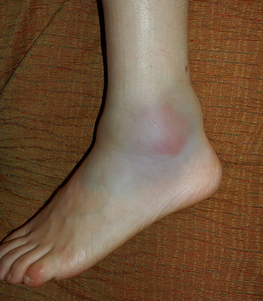
A lateral ankle sprain usually occurs when the foot rolls inward, stretching or tearing the ligaments on the outside of the ankle — most commonly the anterior talofibular ligament (ATFL). It often happens during sudden changes of direction, awkward landings, or stepping on uneven ground.

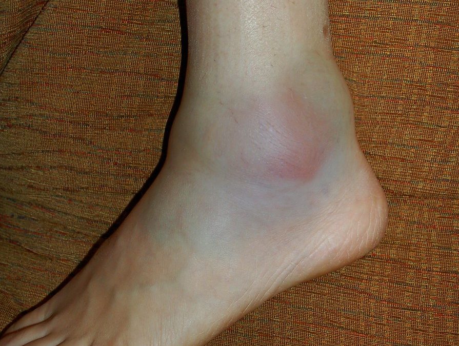
You might feel a pop or crunch, followed by swelling, bruising, and pain when walking or bearing weight. While it may seem like a “simple sprain,” it’s anything but — around 40% of people report long-term issues one year post-injury if not managed well​.

gray concrete statue of a man

Common Mistake: Rest, Ice, and… That’s It?

Too many people still follow the old R.I.C.E. (rest, ice, compression, elevation) model and assume the job is done. While these strategies can help in the first 48 hours, they’re far from sufficient for full recovery.

In fact, research has shown that inadequate rehab is a major contributor to chronic ankle instability — a condition marked by recurrent sprains, feelings of the ankle “giving way,” and reduced confidence in movement​.

CAI can lead to altered biomechanics and poor neuromuscular control, increasing the risk of knee injuries, Achilles tendinopathy, or even hip and low back pain due to compensation.

Proper Rehabilitation Is Key — Here’s What the Evidence Says

Rehabilitation needs to start early and be progressive. High-quality clinical guidelines and systematic reviews strongly support the following strategies:

Functional Support and Early Mobilisation

Functional bracing (like an ankle brace or taping) is preferred over rigid immobilisation and should be used for 4–6 weeks . Early weight-bearing as tolerated leads to quicker return to activity and better outcomes .

Exercise Therapy

Neuromuscular training (balance, proprioception, and strength work) is the foundation of successful rehab. It improves ankle control, prevents recurrence, and reduces the risk of CAI​. A wobble board, single-leg balance, hopping drills, and directional change exercises are all commonly used.

Manual Therapy

Joint mobilisations and soft tissue work may improve dorsiflexion range, decrease pain, and aid in functional recovery​. At Praxis, we combine manual therapy with functional retraining to fast-track performance readiness.

Individualised Return-to-Sport Testing

Return to sport shouldn’t be based on time alone. We use objective testing — including single-leg hop symmetry, balance tests, and strength assessments — to ensure you’re not returning with deficits that could increase your reinjury risk.

The Cost of Incomplete Rehab: What Happens If You Don’t Get It Right?

A rushed or poorly structured rehab may get you back to activity temporarily — but it opens the door to:

  • Chronic Ankle Instability (CAI): Repeated sprains, perceived instability, and loss of ankle confidence.

  • Performance Limitations: Reduced agility, speed, and power due to poor proprioception and strength deficits.

  • New Injuries: Compensatory patterns can lead to medial tibial stress syndrome (shin splints), Achilles overload, or even ACL risk due to poor landing mechanics.

In elite sport, we see this cascade far too often. That’s why our rehab at Praxis isn’t just about the ankle — it’s about restoring whole-limb function and confidence under pressure.

Prevention: Keep Your Ankles Bulletproof

At Praxis Physiotherapy, we don’t just treat ankle sprains — we help prevent them. Our prevention approach includes:

  • Regular Balance and Plyometric Training: Incorporating single-leg exercises into gym and field work.

  • Proprioceptive Work: Using wobble boards, balance mats, and directional hopping.

  • Footwear and Bracing Advice: Particularly for high-risk sports like netball, football, and athletics.

  • Pre-season Screening and Performance Testing: For our affiliated sports clubs and athletic populations.

Evidence supports proprioceptive training as a proven strategy to reduce ankle sprain incidence by up to 35% in high-risk athletes​.

Why Choose Praxis Physiotherapy?

Our exposure to elite sport has taught us what good rehab looks like — and we apply those same high standards to every patient. Our clinics are equipped with strength testing tools, reformer Pilates, and full gym access, giving you the tools to rebuild better.

We understand the mindset of athletes — from juniors chasing state squads to elite-level players returning from surgery. That’s why we tailor your program based on sport demands, movement patterns, and individual goals.

Whether you rolled your ankle playing touch footy or twisted it at work, we’re here to get you back — stronger, faster, and more confident than before.

Need Help with an Ankle Sprain?

If you’ve recently rolled your ankle or are dealing with ongoing instability, book a consultation at Praxis Physiotherapy. Let our team guide you through a structured rehab program grounded in sports science and elite clinical standards.

Until next time, Praxis What You Preach

📍 Clinics in Teneriffe, Buranda, and Carseldine
💪 Trusted by athletes. Backed by evidence. Here for every body.

References

Ruiz-Sánchez et al. (2022). Management and treatment of ankle sprain according to clinical practice guidelines: A PRISMA systematic review. Medicine (Baltimore), 101(42)

Green et al. (2019). What is the quality of clinical practice guidelines for the treatment of acute lateral ankle ligament sprains in adults? BMC Musculoskeletal Disorders, 20(394)

Doherty et al. (2017). Treatment and prevention of acute and recurrent ankle sprain: an overview of systematic reviews with meta-analysis. BJSM, 51(2), 113–125.

Related Articles
Plantar Fasciopathy: Understanding how to heal your heel pain
Feel like your walking on glass in the mornings?  Those first few steps after a long perio…

Shin Splints: Causes, Treatment & How to Get Back to Running Stronger
Key Takeaways Shin splints (MTSS) are an overuse bone stress injury. Training load errors …

Achilles Tendinopathy: How to treat your Achilles Pain

Today on the Praxis What We Preach blog, where we shed light on Achilles tendinopathy, a c…
Plantar Fasciopathy: Understanding how to heal your heel pain

Plantar Fasciopathy: Understanding how to heal your heel pain

Feel like your walking on glass in the mornings?  Those first few steps after a long period of sitting hurt the underside of your heel? Struggling to stand at the end of a long day due to your feet? If so, then you may have plantar fasciopathy, also known as plantar fasciitis. Plantar fasciopathy is a common condition that affects the plantar fascia – a thick band of connective tissue on the bottom of the foot. Plantar fasciopathy commonly affects individuals between the ages of 40 and 60, but can affect almost anyone. In this article, we will delve into the causes, symptoms, treatment options, and preventive measures to help you understand, and more importantly manage, this condition.

Causes and Symptoms

Plantar fasciopathy is often caused by repetitive strain or excessive loading of the plantar fascia, leading to microtears and inflammation. Factors such as overuse, improper footwear, high-impact activities, flat or high-arched feet, and tight calf muscles can contribute to its development. The condition is characterised by sharp pain or a dull ache on the underside of the heel or along the arch of the foot. Pain is typically worse in the morning or after periods of inactivity, and may improve with movement. Standing for long periods or walking on hard floor can also be aggravating.

Treatment Options

The treatment of plantar fasciopathy focuses on reducing pain, promoting load tolerance, and addressing the underlying causes. Physiotherapy interventions play a crucial role in managing this condition. Therapeutic techniques such as manual therapy, stretching exercises, and strengthening exercises can help relieve pain, improve flexibility, and restore foot function. Specifically, improving the windlass mechanism (a phenomena that refers to the tightening of the plantar fascia during the push-off phase of walking or running when you big toe extends). This mechanism helps distribute forces evenly throughout the foot and reduces strain on the plantar fascia. More generally, improvement of the footy intrinsics and plantar flexors more generally have been shown to reduce the severity and duration of symptoms as well.

Additionally, the use of orthotics, taping, or night splints may provide support and alleviate symptoms. Extracorporeal shockwave therapy (ESWT) and ultrasound therapy are also viable treatment options in some cases. In severe or persistent cases, corticosteroid injections or surgery may be considered, though this is usually reserved for when conservative measures have failed.

Preventive Measures

Prevention is key to reducing the risk of plantar fasciopathy starting in the first instance. If you are keen to ‘pound the pavement’ for example, then gradually increase activity levels. Avoid sudden changes in intensity or duration to prevent overloading the foot. This may mean dancing long bouts for the first time in a while, or returning to running post injury. Wear footwear that provides adequate arch support and cushioning. Understand the importance of regular stretching exercises for the calf muscles and plantar fascia.

As physiotherapy professionals, we understand that addressing the symptoms of plantar fasciopathy early is essential for providing effective care. At Praxis, effective care means arming you with adequate advice and education so you can help manage the symptoms yourself. Further, implementing appropriate treatment options and emphasizing preventive measures, we support individuals in overcoming foot pain and restoring quality of life. After all, we aim to Prevent, Prepare, Perform! So if you have heel pain that is stopping you from doing what you would like to do, discuss it with our knowledgeable team today!

Until next time – Praxis What You Preach!

📍 Clinics in Teneriffe, Buranda, and Carseldine

💪 Trusted by athletes. Backed by evidence. Here for everyone.

Related Articles

Achilles Tendinopathy: How to treat your Achilles Pain

Today on the Praxis What We Preach blog, where we shed light on Achilles tendinopathy, a c…

Shin Splints: Causes, Treatment & How to Get Back to Running Stronger

Key Takeaways Shin splints (MTSS) are an overuse bone stress injury. Training load errors …

Ankle Sprains: Don’t Let a Simple Injury Turn into a Long-Term Problem

Ankle sprains are among the most common injuries we see at Praxis Physiotherapy. Whether y…
The Single Leg Squat

The Single Leg Squat

For those of you who have ever read a research article and thought it was a tough read, i’d like to let you in on a little secret. Doing the research is far worse! If only memes were a thing when I started my Masters of Applied Science thesis, I’d have changed my background to remind myself that conducting research was even drier than every dish an apprentice has cooked for Gordon Ramsey.

My mentor at the time, who was the manager of the sports science and sports medicine devision of Cricket Australia and later the head of the AIS human movements department, Dr Marc Portus, enlightened me with something quite profound. He said, “There are two outcomes from a thesis. Either you live it for the rest of you academic days or it sits on a bookshelf for years collecting dust.” Given I completed my thesis and went straight onto my graduate entry physiotherapy masters, it is fair to say i’m in the later camp. Quite a few years have passed now however, so I’d thought i’d dust off the cover and summarise my thesis for all of you playing at home!

So not to degrade my self entirely, I thought I found some pretty good stuff that has affected the way I assess and treat today. I haven’t shared it all today as it was more than 140 pages long and ‘ain’t nobody got time for that’. More importantly though, my thesis reminded me of the passion I have for lower limb biomechanics and that physiotherapy (as opposed to research) was always meant to be on the cards for me. My thesis, “The 3D Kinematics of the Single Leg Flat and Decline Squats” boiled down to looking at how the ankle position changed the joint angles single leg squat, how hip strength affected the squat as well as few other things that aren’t worth mentioning here.

BACKGROUND:

The single leg squat (SLS) replicates an athletic position commonly assumed in sport such as cutting (powerful change in direction while running made from one leg), jumping and balancing which all require the control of the trunk and pelvis on the weight bearing femur in all three planes of movement [1-5].

Photo from the Praxis Physiotherapy article: The Single Leg Squat

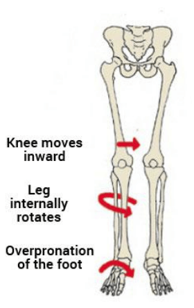
As such, the SLS is commonly used by clinicians as a functional measure of dynamic lumbo-pelvic stability [6-8]. Abnormal movement within the SLS tend to be characterised by the commonly described “medial collapse” or “dynamic valgus”. Specifically, there is excessive femoral internal rotation, femoral adduction, knee valgus, tibial internal rotation and foot pronation of the weight-bearing limb with resultant excursion of the contralateral non weight bearing Ilium and excessive lateral flexion of the trunk [3, 6-8].

The reason why this tends to be perceived as a big deal is that this position tends to be argued as a lack of lumbopelvic stability and results in increased loading of the knee. Moreover, pelvis weakness tends to be ascribed to the absence of stability ultimately resulting in a position in which many acute and overuse injuries of the lower limb may occur. These ailments include, ACL / MCL ruptures, patellofemoral pain syndrome (PFPS), illiotibial band friction syndrome (ITBFS) and shin splints to name a few. That is why the SLS appears to be a valuable rough screening tool in clinical practice.

Photo from the Praxis Physiotherapy article: The Single Leg Squat

MY FINDINGS:

As mentioned, I looked at how a decline board of 20 degrees changed the angles of the lower limb during the squat. I also looked at if any strength measures of the hip related to how someone squatted between conditions. Finally, I looked at if the decline board altered how someone was scored by experienced physiotherapist as a competent or not at the squat

Photo from the Praxis Physiotherapy article: The Single Leg Squat

JOINT ANGLES (KINEMATICS:)

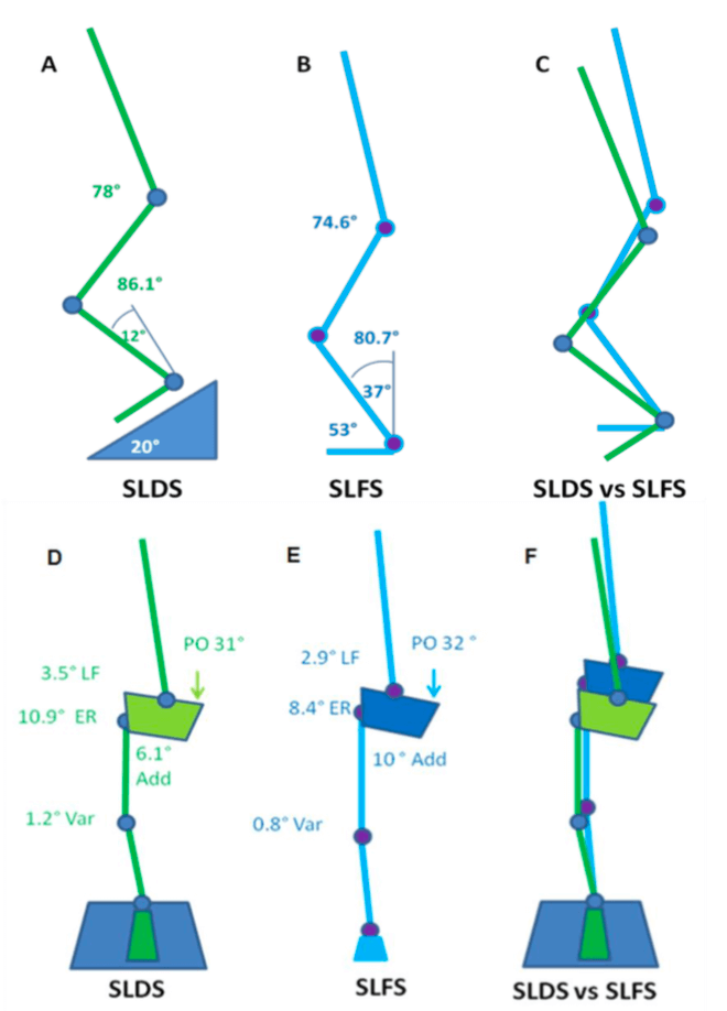
A picture tells a thousand words so in the interests of brevity, the stick squat figure is essentially a summary of two years of work.

So what this means, when someone performs a SLS on a flat surface, relative to a decline surface they tend to have:

  • A more upright torso
  • More rotation of the pelvis toward the weigh bearing (WB) limb
  • Reduced flexion but more adduction and internal rotation of the thigh on the WB hip (pelvic close to femur)
  • Less flexion of the knee but the same position relative to the foot as you look from the front (known as frontal plane knee excursion) at the bottom of range
  • Reduced internal rotation of the shin
  • Reduced ankle flexion

Essentially, in a flat squat you tend to ‘corkscrew’ your pelvis and adopt the medial collapse position much more easily than in the decline squat position. This may because of ankle range of motion issues as well as the ability to adequately recruit pelvic musculature. Yep – two years to get that!

REVISITING THE DATA (2026 RE-ANALYSIS):

Since I first wrote this piece, I have gone back to the original 3D motion capture dataset from my Masters and re-analysed it using more rigorous statistics than I had access to at the time. This work is unpublished and currently being prepared for peer review, so treat it as “watch this space” rather than settled science, but it is a meaningful sharpening of the original findings and, in one respect, a genuine correction to what I wrote above.

The original analysis compared single joint angles at one point in the squat. The re-analysis instead used a multivariate test (Hotelling’s T²) across five hip and pelvis variables together, a false-discovery-rate corrected sweep across eleven segments of the entire kinetic chain (ankle to thorax), and a continuous waveform comparison across the full 0-100% squat cycle rather than a single snapshot.

Two things held up, and one thing did not.

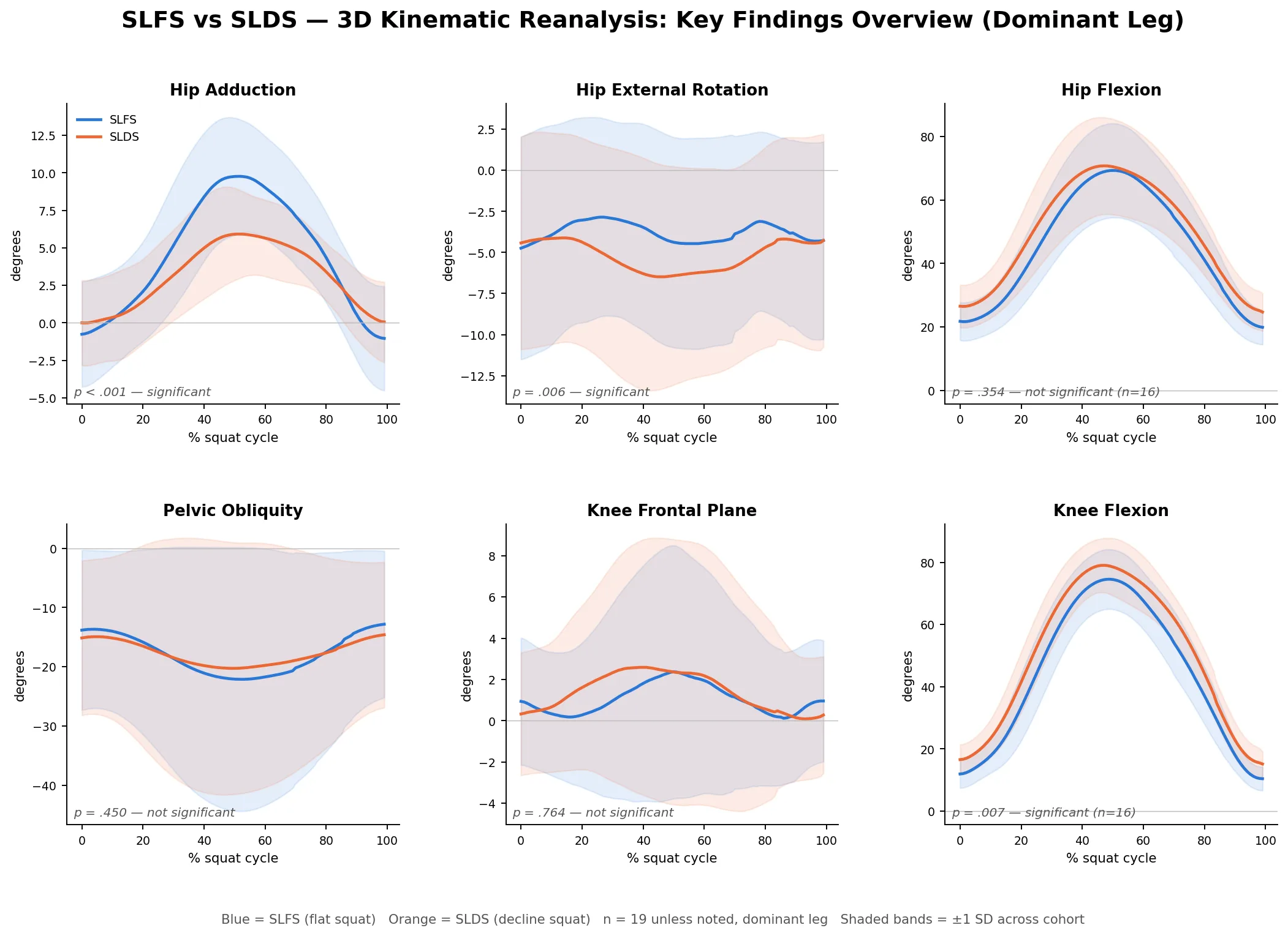
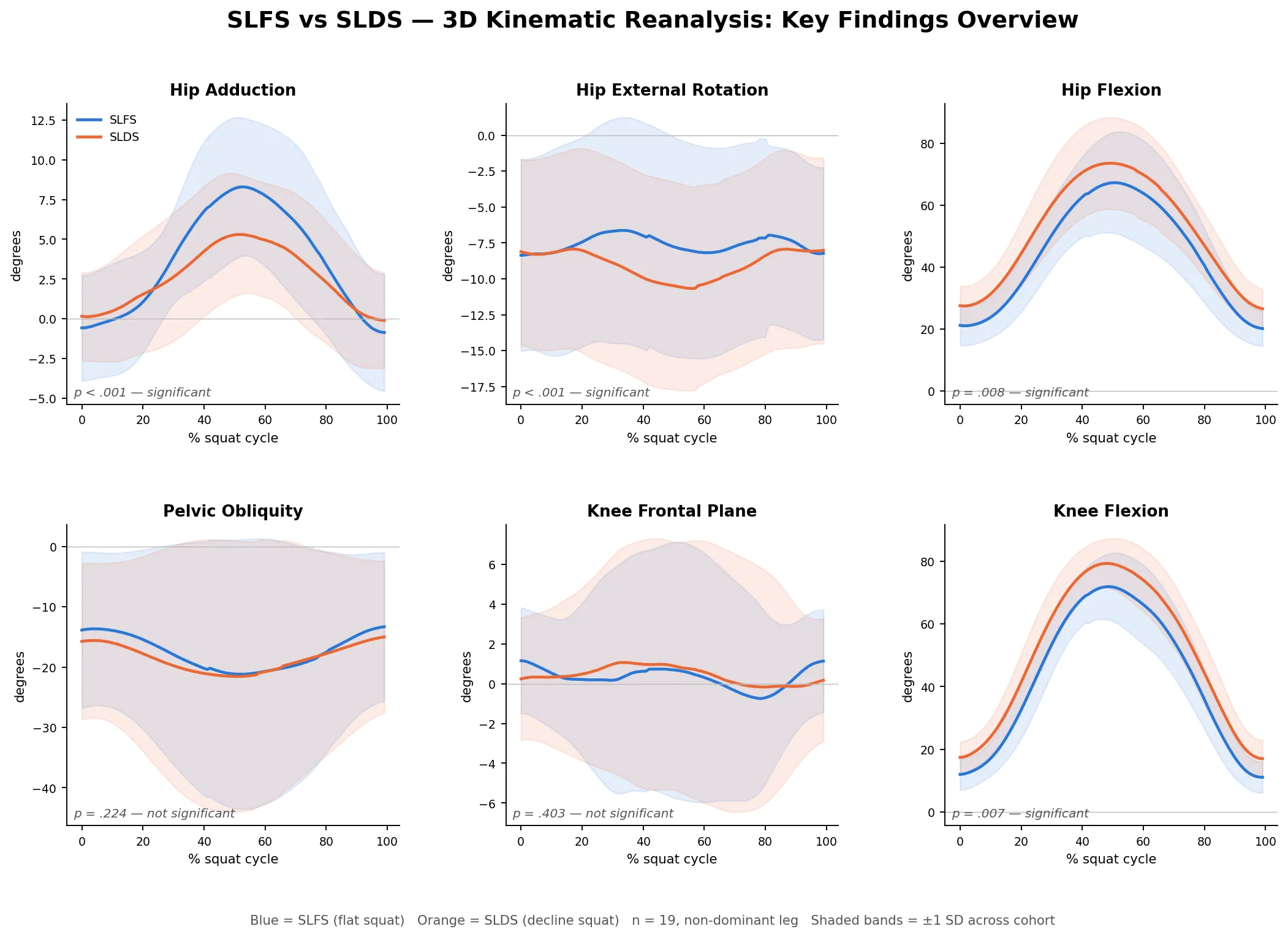
What held up: hip adduction and hip external rotation were consistently greater on the flat squat than the decline squat, by roughly 2-4 degrees on average, and this was robust to the multivariate correction, not just a single-variable finding. Of the eleven segments tested across the whole kinetic chain, only the weight-bearing ankle and the weight-bearing hip reliably differed between conditions (both p<0.001 after FDR correction) - the pelvis, lumbar spine and thorax did not differ. The continuous waveform data also showed that this divergence is not constant through the squat - it emerges gradually through descent and peaks near the deepest point, then closes again on the way up, which matters if you are assessing someone visually and only glance at the very bottom of the movement.

What did not hold up: the “medial collapse” / frontal-plane knee position I described above, comparing flat to decline squat, was not significantly different between the two conditions in this more rigorous analysis. The knee did move differently between the two squat variations, but the difference was in the sagittal (flexion) and transverse (rotation) planes, not the frontal plane. In plain terms, if you are judging flat-versus-decline squat technique by eye and using “does the knee cave in” as your marker, this data suggests that is not actually a variable that distinguishes the two techniques, even though the knee genuinely is moving differently between them in other planes.

Two figures below summarise this: continuous waveform plots (mean ± 1 SD) across the squat cycle for hip adduction, hip external rotation, hip flexion, pelvic obliquity, frontal-plane knee position and knee flexion, for both the dominant and non-dominant limb.

Cohort-averaged 3D kinematic waveform plots across the squat cycle for the dominant limb: hip adduction, hip external rotation, hip flexion, pelvic obliquity, frontal-plane knee position and knee flexion, comparing single leg flat squat and single leg decline squat

Figure 1: Dominant leg, n=19.

Cohort-averaged 3D kinematic waveform plots across the squat cycle for the non-dominant limb: hip adduction, hip external rotation, hip flexion, pelvic obliquity, frontal-plane knee position and knee flexion, comparing single leg flat squat and single leg decline squat

Figure 2: Non-dominant leg, n=19.

A couple of honest caveats: this dataset has no force plate data, so none of this is a statement about joint torque, only joint angle. Limb dominance was self-reported (kicking leg), not independently tested, and squat depth and tempo were self-selected rather than standardised between conditions. I’d treat this as a more careful re-reading of the same movement pattern, not a wholesale rewrite of the original conclusion – the ankle-hip relationship I originally described still stands up, it’s just the frontal-plane knee story that needs dropping.

STRENGTH AND MOVEMENT:

My results demonstrated a tendency for the pelvis to remain increasingly level with greater hip abduction strength. However, the relationship between strength and the pelvis was observed in the decline condition but not the flat condition. This may be due to hip abduction was shown to be significantly less (more neutral) in the SLDS which seemingly promoted greater muscle activation and subsequent control of pelvis. The self selection of squat depth may have also been a critical factor in finding as those with weak hips may have squatted deep to adopt maladaptive positions. Previous research has indicated that the hip abductors and external rotators play an important role in lower extremity alignment as they assist in the maintenance of a level pelvis [9] and are capable in balancing a number of biomechanical forces in the body [10].

Interestingly, there were no significant relationships observed between hip abduction strength and knee valgus (knee falling in) for both squatting conditions. There was however a trend between hip abduction strength and knee valgus which supported previous research. It is keeping with the assumption that increased knee valgus might also be associated with reduced hip abduction and external rotation strength [11].

SUMMARY:

  • To maximise athletic function, particularly in sports such as soccer, netball and AFL, stability through the pelvis and hips, proximal lower limb, spine and abdominal structures is required [12].
  • The importance of pelvis stabilisation for lower extremity injury prevention [13] particularly the knee [14-17] has been well documented in the literature.
  • Adequate lumbopelvic-femur strength and muscle function may conceivably reduce exposure to other intrinsic risk factors such as inefficient force attenuation, unstable movement patterns and lower limb malalignments during activity [18, 19].
  • Ankle flexibility may also be a factor in lower limb physical resilience and injury prevention.
  • Support for the previous statements has been demonstrated in the relationships between hip strength measures and kinematics within selected results of my study.

There you have it. Two years of my life summarised to a few paragraphs. From a personal perspective, I took away from my research experience to be always questioning why we do things and see if there is someone out there who has answered the questions we seek. Finally, don’t overcook chicken – Ramsay doesn’t like it.

Until next time, Praxis What You Preach

📍 Clinics in Teneriffe, Buranda, and Carseldine

💪 Trusted by athletes. Backed by evidence. Here for everyone.

Related Articles

Split Squat vs Squat vs Deadlift: How to tailor your lower body training

The age old question: What’s the best gym activity for my sport? Well – the an…

Unilateral vs Bilateral Training – Part 2: Performance outcomes

In part 1 of this blog, we discussed the generalities associated with bilateral vs unilate…

Unilateral vs Bilateral Training: Part 1 – Your Comparative Guide

We often get asked what are the benefits of one form of exercise versus another. Sometimes…

References

  1. Neely, F.G., Intrinsic risk factors for exercise-related lower limb injuries. Journal of Sports Medicine, 1998. 26(4): p. 253-263.
  2. Parkkari, J., U.M. Kujala, and K. Pekka, Is it possible to prevent sports injuries? Review of controlled clinical trials and recommendations for future work. Sports Medicine, 2001. 31(14): p. 985-995.
  3. Lysens, R.J., et al., The accident -prone and overuse-prone profiles of the young athlete. The American Journal of Sports Medicine, 1989. 17(5): p. 612-619.
  4. Egger, G., Sports injuries in Australia: causes, costs and prevention. A report to the national better health program., ed. C.f.H.P.a. Research. 1990, Sydney.
  5. Orchard, J.W. and C.F. Finch, Australia needs to follow New Zealand’s lead on sports injuries. The Medical Journal of Australia, 2002. 177: p. 38-39.
  6. Wu, G. and P.R. Cavanagh, ISB recommendations for standardization in the reporting of kinematic data. Journal of Biomechanics, 1995. 28: p. 1257- 1261.
  7. Siegal, P., R. Brackbill, and G. Heath, The epidemiology of walking exercise: implications for promoting activity among sedentary groups. American Journal of Public Health, 1995. 85(5): p. 706-710.
  8. Nicholl, J.P., P. Coleman, and B.T. Williams, The epidemiology of sports and exercise related injury in the United Kingdom. British Journal of Sports Medicine, 1995. 29(4): p. 232-238.
  9. Burnet, E.N. and P.E. Pidcoe, Isometric gluteus medius muscle torque and frontal plane pelvic motion during running. Journal of Sports Science and Medicine, 2009. 8: p. 284-288
  10. Niemuth, P., et al., Hip muscle weakness and overuse injuries in recreational runners. Clinical Journal of Sports Medicine, 2005. 15(1): p. 14-21.
  11. Hollman, J.H., et al., Relationships between knee valgus, hip-muscle strength, and hip-muscle recruitment during a single-limb step down. Journal of Sport Rehabilitation, 2009. 18: p. 104-117.
  12. Kibler, W.B., J. Press, and A. Sciascia, The role of core stability in the athletic function Journal of Sports Medicine, 2006. 36(3): p. 189-198.
  13. Leetun, D.T., et al., Core stability measures as risk factors for lower extremity injury in athletes. Medicine & Science in Sports & Exercise, 2004. 36(6): p. 926-934.
  14. Cichanowski, H., et al., Hip strength in collegiate female athletes with patellofemoral pain. Medicine & Science in Sport & Exercise, 2007. 39(8): p. 1227-1232.
  15. Ireland, M.L., et al., Hip strength measures in female with and without patellofemoral pain. Journal of Orthopaedic & Sports Physical Therapy, 2003. 33(11): p. 671-676.
  16. Nicholas, J.A., A.M. Strizak, and G. Veras, A study of thigh muscle weakness in different pathological states of the lower extremity. American Journal of Sports Medicine, 1976. 4: p. 241-248.
  17. Prins, M.R. and P.V.D. Wurff, Females with patellofemoral pain syndrome have weak hip muscles: a systematic review. Australian Journal of Physiotherapy, 2009. 55: p. 9-15.
  18. Willson, J.D., M.L. Ireland, and I. Davis, Core strength and lower extremity alignment during single leg squats. Medicine & Science in Sports & Exercise, 2006. 38(5): p. 945-952.
  19. Lee, D., The pelvic girdle: An approach to the examination and treatment of the lumbopelvic-hip region. 3rd ed. 2004, Edinburugh: Churchill Livingston.