Navigating Knee Osteoarthritis: A Physio-Centric Pathway to Strength and Mobility Before Surgery

Navigating Knee Osteoarthritis: A Physio-Centric Pathway to Strength and Mobility Before Surgery

Knee osteoarthritis (OA) is one of the most common causes of chronic pain and mobility restriction in Australians over 45. Whether you’re a weekend warrior, an active grandparent, or someone just trying to keep up with the daily demands of life, OA can slowly erode your confidence in movement — long before X-rays show the full extent of joint degeneration.

At Praxis Physiotherapy, we take a forward-thinking, collaborative approach to managing knee OA. Working closely with renowned orthopaedic knee surgeon Dr. Kelly Macgroarty and drawing from our extensive experience with high-performance athletes and everyday patients alike, we believe the journey toward better knees starts well before surgery — and, for many, might even avoid or delay it altogether.

What is Knee Osteoarthritis?

Knee OA is a progressive condition involving the breakdown of joint cartilage and underlying bone, typically leading to:

  • Pain during or after activity

  • Morning stiffness or stiffness after rest

  • Swelling and inflammation

  • Loss of flexibility and range of motion

  • Difficulty with stairs, kneeling, or prolonged standing

Radiographic OA becomes more common with age, but symptoms often precede visible changes on X-ray. Up to 30% of people over 65 show radiographic OA, yet many remain functionally capable — highlighting the importance of early, movement-based interventions (Naja et al., 2021).

Why a Physio-Led Model Before Knee Replacement?

Surgery is not the first or only option. A large systematic review of 19 randomized controlled trials found that non-surgical interventions such as physiotherapy, platelet-rich plasma (PRP), and structured exercise were associated with meaningful improvements in pain and function over 12 months (Naja et al., 2021). Physiotherapy, in particular, is consistently supported by international guidelines as a first-line treatment (Fransen et al., 2015; Bennell et al., 2014).

Traditionally, knee OA rehab has emphasised quadriceps strengthening — and for good reason, as quadriceps weakness is strongly linked to OA-related pain and disability. However, more recent research suggests that focusing exclusively on the quadriceps may be too narrow. Programs that include hip (gluteal), hamstring, and calf muscle strengthening are now shown to be superior in improving functional outcomes, especially for activities like walking, stair climbing, and maintaining balance (Bennell et al., 2014). This broader approach addresses the full kinetic chain around the knee, optimises joint load distribution, and better supports long-term movement efficiency.

At Praxis, our physios:

  • Assess gait, strength, joint mobility, and function

  • Design individualised exercise programs targeting quadriceps, glutes, and calf strength

  • Implement manual therapy techniques to restore joint mobility

  • Provide pain education, load management advice, and real-world strategies

  • Monitor progress and adjust programs over time

This proactive approach not only builds resilience in the knee but also prepares the joint and surrounding muscles should surgery eventually be required.

Booster Sessions: Keeping Gains, Lowering Costs

An often-overlooked strategy is the use of booster physiotherapy sessions — structured follow-ups after an initial rehab program. Research by Bove et al. (2018) showed that exercise programs with booster sessions at 3, 6, and 12 months were not only more clinically effective but also more cost-effective over a two-year period compared to standard physiotherapy care.

At Praxis, we now embed these booster sessions into long-term OA management. They help patients:

  • Maintain strength and conditioning gains

  • Stay accountable with home programs

  • Troubleshoot new symptoms early

  • Reduce future health care costs and medication reliance

What About Injections and Other Adjuncts?

We often collaborate with GPs and orthopaedic specialists to incorporate adjunct treatments where the evidence supports it:

  • Platelet-rich plasma (PRP) injections showed significant long-term benefit for pain and function, with improvements of ~20 points on the WOMAC index. PRP ranked just behind stem cells as the most effective non-surgical treatment in a large 2021 network meta-analysis (Naja et al., 2021).

  • Hyaluronic acid (HA) injections have shown mixed results. A review of overlapping meta-analyses concluded that HA is likely safe and modestly effective, especially in early-stage OA, although guideline recommendations remain inconsistent (Xing et al., 2016).

Ultimately, our philosophy is to build strong knees first, and complement physiotherapy with interventions like PRP or HA only when clinically indicated and appropriately timed.

Surgical Collaboration 

In more advanced cases, where conservative management fails, we work closely with Dr. Kelly Macgroarty, one of Queensland’s leading knee surgeons. Our relationship allows:

  • Streamlined triage for surgical consultation

  • Shared prehabilitation planning to improve surgical outcomes

  • Integrated post-operative rehab, using in-clinic gym equipment and reformer Pilates to accelerate return to function

This continuity ensures you’re never left navigating knee OA alone — whether your journey stays entirely within physio care or progresses to surgical management.

Why Praxis Physiotherapy?

At Praxis, we’ve built our care model around best-practice guidelines, decades of elite sport and private practice experience, and a shared goal of keeping our patients active, independent, and thriving.

Our Teneriffe, Carseldine and Buranda clinics offer:

  • In-clinic rehab gyms

  • Reformer Pilates for joint-friendly loading

  • Real-time strength testing technology

  • Physios with elite sports and post-surgical rehab experience

Take the First Step

If you or someone you love has been told you’re “heading for a knee replacement,” don’t wait. There is so much we can do to reduce pain, improve function, and build confidence in your knees — surgery or not.

Book an appointment today at one of our Brisbane clinics and start your journey to stronger, more resilient knees.

Interested in ACL specific rehab? Check our guide on return to sport after ACL injury

Until next time, Praxis What You Preach!

📍 Clinics in Teneriffe, Buranda, and Carseldine

💪 Trusted by athletes. Backed by evidence. Here for everyone.

References

  • Bove, A. M., Smith, K. J., Bise, C. G., et al. (2018). Exercise, manual therapy, and booster sessions in knee osteoarthritis: cost-effectiveness analysis from a multicenter randomized controlled trial. Physical Therapy, 98(1), 16–27.

  • Fransen, M., McConnell, S., Harmer, A. R., Van der Esch, M., Simic, M., & Bennell, K. L. (2015). Exercise for osteoarthritis of the knee: a Cochrane systematic review. British Journal of Sports Medicine, 49(24), 1554–1557.

  • Bennell, K. L., Dobson, F., & Hinman, R. S. (2014). Exercise in osteoarthritis: moving from prescription to adherence. Best Practice & Research Clinical Rheumatology, 28(1), 93–117.

  • Naja, M., Fernandez De Grado, G., Favreau, H., et al. (2021). Comparative effectiveness of non-surgical interventions in the treatment of patients with knee osteoarthritis: a PRISMA-compliant systematic review and network meta-analysis. Medicine, 100(49), 

  • Xing, D., Wang, B., Liu, Q., et al. (2016). Intra-articular hyaluronic acid in treating knee osteoarthritis: a PRISMA-compliant systematic review of overlapping meta-analyses. Scientific Reports, 6, 32790.

Fact or Fiction Friday – Lower back Pain and MRI’s

Fact or Fiction Friday – Lower back Pain and MRI’s

I need to get an MRI to help with the management of my lower back pain

Answer – FICTION

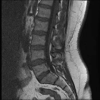
In a recent narrative review, Wang and colleagues (2018) concluded that MRI imaging in the early stages of lower back pain can have detrimental effects including more pain, less improvement, higher risk of surgery and worse overall health status. In fact, one study reported that patients that received an MRI within the first month had an 8x greater risk for surgery and 5x more medical costs!

If you do NOT present with severe neurological deficits, signs of a serious or specific underlying condition or have persistent pain >6 weeks which is unresponsive to conservative treatment then there likely isn’t a need for further investigation!

To get help with your long standing back pain or even that acute flare up, give us a call on (07) 3102 3337 or book online  so we can sort you out.

#praxiswhatyoupreach #praxisphysio #factorfictionfriday #physioeducation #preventprepareperform #pain #backpain #lowerbackpain #MRI #patienteducation

Wang Y, et al. Informed appropriate imaging for low back pain management: A narrative review. Journal of Orthopaedic Translation. 2018.

FACT OR FICTION FRIDAY || All rotator cuffs tears need surgery.

FACT OR FICTION FRIDAY || All rotator cuffs tears need surgery.

Answer: FICTION. The devil is in the detail!

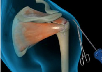
The rotator cuff is a group of 4 muscles that aid in providing stability through range for the shoulder joint, particularly overhead. There are multiple risk factors for RC tears, but most are down to overactivity of the shoulder joint decreased conditioning of the shoulder complex, which comes with age. In fact, cuff tears are common in individuals over the age of 40 with linear increase in incidence as we get older.

Pain with movement and function is one of the biggest symptoms of a rotator cuff tear. However, it is important to know that a sizeable portion of RC tears are actually asymptomatic and don’t cause the person any pain or discomfort! A study by Minagawa and Yamamoto in 2013 found that in a screening of 664 village residents, 147 subjects had RC tears on a medical imaging screening. Surprisingly 65% of them had no symptoms at all and didn’t have any shoulder complaints.

Mid Potion Achilles Tendinopathy Location

The two main ways of treating a cuff tear is either through conservative management with your physiotherapist or down the surgical route, which is also then followed by physiotherapy rehabilitation.

SO, back to the original question: “Do I need surgery?”. As always – it is a case by case decision!! There is no definitive evidence for supporting one over the other generally speaking!

However, it is vitally important to note that each option comes with their own pros and cons. Furthermore, it is important to remember that just as every person is different, each case of rotator cuff tear is different. Young vs old, acute vs degenerative RC tear, current and desired future function, pain levels, radiographic findings, previous history of shoulder trauma and the patient’s wishes are only some of the questions that aid in the decision process.

The best way to decide would be to contact us to asses you and discuss both options and what your goals of rehab are so that a tailored plan can be developed WITH you. To read more about RC tears, read our blog here

To get your shoulder back on track, book online or give us a call on (07) 3102 3337.

Team Praxis

PREVENT | PREPARE | PERFORM

Stingers AKA Neural traction injuries

Stingers AKA Neural traction injuries

SUMMARY:

  • Stingers are essentially a tractioning of the neural system
  • This can cause pain, movement and sensation changes
  • Chronic traction to the nervous system can have a cumulative effect on nerve function
  • The more damage to the nerve, the more serious the outcome
  • We shouldn’t be as dismissive of “stingers”, particularly if they are recurrent
  • Physiotherapy has a role to play

STINGERS:

Stingers have been the catch cry of many contact sports over the years. Often dismissed as ‘just a stinger’, trauma to the nervous system should probably be taken a little more seriously, as we delve into detail today.

Stingers are most commonly experienced in contact sports whereby the shoulder of a players is forcefully depressed, as experienced with a tackle in NRL or union. A large range of motion over a short period of time can result in a ‘traction’ of the Brachial Plexus (a network of nerves formed from exiting branches of the spinal cord in the neck that transverse to the shoulder and arm). Thus network of nerves sends signals from your spinal cord to your shoulder, arm and hand and thereby providing feeling and movement to these regions.

BACKGROUND:

To understand a stinger injury, a clear understanding of nerves and nerve related injuries is required. A nerve is an enclosed, cable-like bundle of nerve fibres called axons, in the peripheral nervous system. A nerve provides a common pathway for the electrochemical nerve impulses that provide a number of functions, including getting our muscles to move!

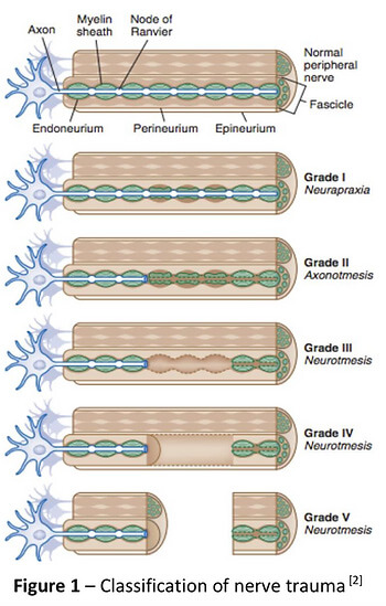
Seddon and Sunderland present a five-grade classification scale for nerve related injuries[1-4]. Figure 1 illustrates the physiological changes that occur through each grade of injury. Essentially it outlines the greater amount of disruption to the anatomy of the nerve.

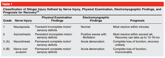
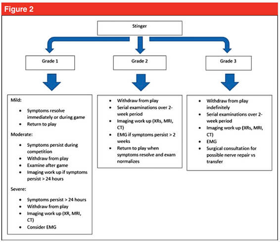
The more damage to the nerve, the more serious the outcome. Table 1 outlines the three different grades of stinger injuries.[4, 5] The most common stinger is a grade 1 injury, which represents a neurapraxia, or nerve stretch injury, without axonal disruption.[4] In an acute setting, this can result in motor and sensory loss/changes, which usually resolve within minutes.[1, 4] Grade 2 and 3 involve a higher degree of nerve injury, usually involving a crush, transection or compression mechanism.[1]

Chronic traction to the nervous system can have a cumulative effect on nerve function. This is termed “chronic stinger syndrome” and represents a distinct entity from acute stingers that may reflect long- standing structural changes of the subaxial spinal canal and chronic irritation/degeneration of the exiting nerve root complex.[4, 6]. This sounds complex but essentially means

Mid Potion Achilles Tendinopathy Location

A Clinical Example from Zac

“During a Gridiron match, a player was injured whilst making a tackle. I reviewed the player on field and he was unable to utilise his right upper limb (full paralysis) from shoulder down to his hand. The player was removed from the field immediately to be further assessed and monitored. A complete neurovascular assessment was performed, assessing motor function/strength, sensation, reflexes, and vascular status, as well was the cervical spine. Motor and sensation changes were the only deficits noted and were reviewed frequently. After roughly five minutes, the player demonstrated full upper limb motor strength and sensation, with nil lingering symptoms. In collaboration with the patient, it was decided he would return to match play immediately. The patient was monitored throughout the game and reported no further symptoms.”

Figure 2 shows a proposed decision tree when managing stinger injuries.[4] This clinical example outline above fits the Grade 1 Mild category as he was able to return to competition with nil lingering symptoms. Despite the lack of symptoms during the game, it is recommended the patient be reviewed again both after the game and weekly for two weeks to ensure a full resolution of symptoms.[4, 7]

The role for neural mobilisation?

Current non-surgical management involves rest, pain control and resistance training[4]. Though not explored within the literature, neural mobilization may have an important role in patients with persistent symptoms, such as Grade 1 moderate to severe, and more recurrent neuropraxias. Though not assessed in this specific population, there is evidence for neural tissue management being superior to minimal intervention for pain relief and reduction of disability in nerve related chronic musculoskeletal pain.[8] It is biologically plausible that recurrent neuropraxias may respond in a similar way, utilising neural mobilisation (tensioning or sliding) and mobilisation of surrounding structures.

Management of persistent Grade 1 injuries may differ slightly, specifically if the suspected mechanism of injury was through traction rather than compression. The nerve structures may have a heightened sensitivity to tensioning based techniques due to the similar mechanism of injury and may respond better acutely to sliding techniques which limit the strain on the nerve and focus on excursion. Tensioning techniques may be important in the sub-acute phase by loading the patient’s nervous system (i.e. increased strain) in preparation for return to function (i.e. tackling with acute traction on the brachial plexus).

In summary, perhaps we shouldn’t be as dismissive of “stingers”, particularly if they are recurrent for you! If you have any questions or would like to see one of our physios regarding your injury, feel free to contact us on (07) 3102 3337 or book online on our website

Till next time, Praxis what you Preach

Team Praxis

Prevent | Prepare | Perform

REFERENCES:

Menorca, R.M.G., T.S. Fussell, and J.C. Elfar, Nerve physiology: mechanisms of injury and recovery. Hand clinics, 2013. 29(3): p. 317-330.

Tsao B, B.N., Bethoux F, Murray B, Trauma of the Nervous System, Peripheral Nerve Trauma. 6th ed. In: Daroff: Bradley’s Neurology in Clinical Practice. 2012.

Sunderland, S., A classification of peripheral nerve injuries producing loss of function. Brain, 1951. 74(4): p. 491-516.

Ahearn, B.M., H.M. Starr, and J.G. Seiler, Traumatic Brachial Plexopathy in Athletes: Current Concepts for Diagnosis and Management of Stingers. J Am Acad Orthop Surg, 2019.

Feinberg, J.H., Burners and stingers. Phys Med Rehabil Clin N Am, 2000. 11(4): p. 771-84.

Presciutti, S.M., et al., Mean subaxial space available for the cord index as a novel method of measuring cervical spine geometry to predict the chronic stinger syndrome in American football players. J Neurosurg Spine, 2009. 11(3): p. 264-71.

Aldridge, J.W., et al., Nerve entrapment in athletes. Clin Sports Med, 2001. 20(1): p. 95-122.

Su, Y. and E.C. Lim, Does Evidence Support the Use of Neural Tissue Management to Reduce Pain and Disability in Nerve-related Chronic Musculoskeletal Pain?: A Systematic Review With Meta-Analysis. Clin J Pain, 2016. 32(11): p. 991-1004.

FACT OR FICTION FRIDAY || I’m too old to lift weights!

FACT OR FICTION FRIDAY || I’m too old to lift weights!

Answer: FICTION 🙊 Progressive strength training in the elderly (>60 years) is efficient, even with higher intensities, to improve bone health, pack on muscle and retain function. And not surprisingly, side effects are rare! Strength training increases muscle strength by increasing muscle mass, and by improving the recruitment of motor units, and increasing their firing rate. This is no different between younger and older gym goers.
Mid Potion Achilles Tendinopathy Location

It all comes down to how you train! Training with higher loads generally provokes marginally larger gains in muscle size. Intensity corresponding above 85% of the individual maximum voluntary strength can also illicit improved rate of force development compared to 60-80%. This is imperative for reducing frailty as we age.

It is now recommended that healthy old people should train 3 or 4 times weekly for the best results; persons with poor performance at the outset can achieve improvement even with less frequent training.

So if you are using age as an excuse – STOP! Don’t let your age be a barrier to trying new things or feeling strong. We are here to help with supervised sessions, a great network of PTs as well as our clinical reformer pilates classes which are a great way to start (or return) to strength training!

To book for a clinical pilates 1:1 session or to chat with a physio about how strength training can help you, head to our booking page or give us a call on (07) 3102 3337

Team Praxis

PREVENT | PREPARE | PERFORM

References:

Mayer, F., Scharhag-Rosenberger, F., Carlsohn, A., Cassel, M., Müller, S., & Scharhag, J. (2011). The intensity and effects of strength training in the elderly. Deutsches Ärzteblatt International, 108(21), 359.

Lopez, P., Pinto, R. S., Radaelli, R., Rech, A., Grazioli, R., Izquierdo, M., & Cadore, E. L. (2018). Benefits of resistance training in physically frail elderly: a systematic review. Aging clinical and experimental research, 30(8), 889-899.