ACL Reconstruction Rehab – Week-by-Week Recovery Guide with Praxis Physio
Overview
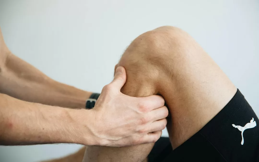
ACL reconstruction surgery marks the beginning of a structured rehabilitation journey. At Praxis Physiotherapy, located in Teneriffe, Carseldine, and Buranda, we provide an evidence-based approach to guide patients from surgery to sport. Backed by over a decade of experience with football teams across Brisbane and collaborative ties with local knee surgeons, our programs are scientifically informed and results-driven.
Research has shown that a phased, criterion-based rehab plan reduces complication rates and improves return-to-sport outcomes (Shelbourne & Nitz, 1990). The following week-by-week overview reflects current best practice from leading ACL rehab literature.
Week-by-Week ACL Rehab Milestones
Prehab: Starting Strong Before Surgery
If you’re waiting for ACL surgery and your knee has no complicating factors like meniscal locking, there’s good evidence that doing some early rehab — before going under the knife — can significantly improve your recovery trajectory. This phase, often called “prehab,” aims to reduce swelling, restore full knee extension, activate the quadriceps, and build general lower limb strength.
Research shows that patients who enter surgery with better quadriceps strength and full range of motion recover faster and regain function more effectively post-operatively (Eitzen et al., 2010). In fact, one study in the British Journal of Sports Medicine found that even just 5 sessions of targeted prehab improved early post-op outcomes like walking speed, strength, and self-reported function (Failla et al., 2016).
Weeks 0–2: Pain, Protection, and Range
Early rehabilitation begins with swelling and pain management, protection of the graft, and restoration of knee extension. Controlled range-of-motion (ROM) exercises and quadriceps activation, particularly of the vastus medialis, are prioritised. Patients often use crutches to maintain safe gait patterns. Early introduction of blood flow restriction (BFR) training supports muscle maintenance without joint overload (Zazirnyi et al., 2020).
Checkpoint: Achieve full extension and minimal swelling by Week 2.
Weeks 2–6: Regain Motion and Begin Strength
Once inflammation is controlled, attention shifts to regaining full ROM, normalising walking gait, and initiating basic strength exercises such as mini-squats and heel raises. Use of closed kinetic chain exercises is supported for their functional benefit and reduced joint stress (Awad et al., 2017).
Checkpoint: Full ROM with independent walking and neuromuscular control.
Weeks 6–12: Strength Foundation
Patients now begin progressive resistance training using clinic gym equipment, including leg presses, Romanian deadlifts, and lunges. Core strength and dynamic control are emphasised. Light cardio via cycling or elliptical may be introduced. Pilates reformers are utilised at Praxis for controlled joint loading and core development.
Checkpoint: Strength symmetry reaching 70% of non-injured leg; competent single-leg stance.
Weeks 12–20: Power and Plyometric Preparation
This phase involves development of reactive strength and neuromuscular readiness. Jump landing, eccentric hamstring training, and lateral movement patterns are introduced. Key focus is on building capacity for eventual cutting and pivoting movements.
Checkpoint: Successful hop tests, 80% limb symmetry, and controlled change-of-direction drills.
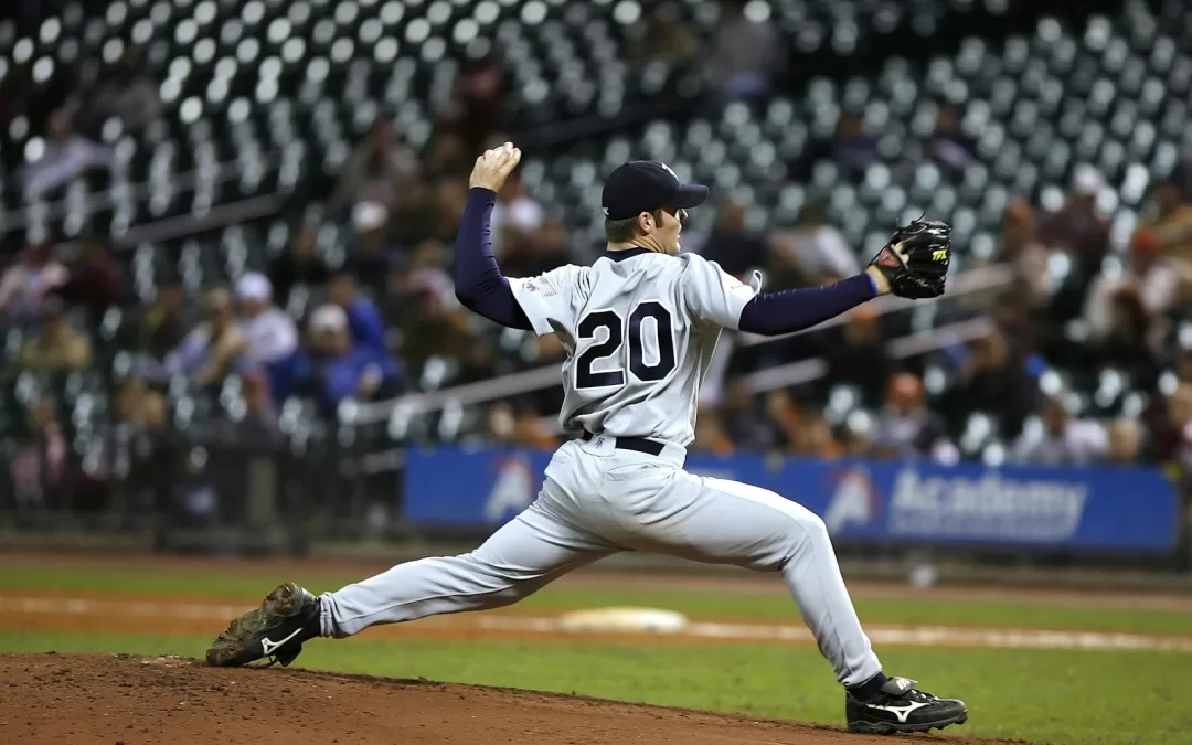
Weeks 20–36: Agility and Functional Sport Movements
Higher-level drills simulate sport-specific movements. Patients perform acceleration/deceleration tasks, direction changes, and reactive decision-making. Plyometrics are progressed in intensity and volume. According to Damian & Damian (2018), phase-specific drills improve psychological readiness and functional return to play (Damian & Damian, 2018).
Checkpoint: Limb symmetry >90% in strength and hop metrics.
Months 9–12: Return-to-Sport Preparation
This stage addresses psychological readiness and simulates sport-specific loading. Functional and fatigue testing are conducted, often including contact drills. Clearance depends on achieving objective strength and control measures (Shelbourne & Patel, 1996).
Checkpoint: Refer for return-to-sport testing (detailed in a separate blog).
Practical Insights for Patients
ACL rehab can be a long and often isolating journey. Many patients report psychological challenges, especially during the early and middle stages when progress may feel slow. At Praxis, we recognise that motivation is a vital part of recovery. Our goal isn’t just to return you to pre-injury levels — it’s to help you become a more resilient, stronger athlete than before. Many ACL injuries stem from non-contact mechanisms linked to strength, control, and movement quality. By targeting these factors throughout rehab, we aim to reduce reinjury risk and elevate athletic performance in the long term.
ACL rehabilitation is about restoring whole-body function, not just healing a ligament. At Praxis Physiotherapy, we use a combination of BFR, Pilates reformers, and in-house rehab gyms to deliver tailored and progressive programs. Our partnerships with Brisbane’s knee specialists ensure timely updates and coordinated care transitions.
Why Brisbane Athletes Choose Praxis Physiotherapy
- Proven protocols developed with elite Brisbane football teams
- Direct lines of communication with top knee surgeons
- BFR and Pilates integrated into post-op care
- Ongoing strength and functional assessments across all phases
Conclusion
A structured, evidence-backed approach to ACL rehab dramatically improves recovery outcomes. At Praxis Physiotherapy, we offer a seamless, week-by-week program from day one post-op through to full sport readiness. Our comprehensive model ensures that Brisbane athletes receive the highest standard of care at every stage. If you’re ready to get your rehab underway, book with us!
Until next time – Praxis What You Preach
📍 Clinics in Teneriffe, Buranda, and Carseldine
💪 Trusted by athletes. Backed by evidence. Here for everyone.
References
- Shelbourne, K.D., & Nitz, P. (1990). Accelerated rehabilitation after anterior cruciate ligament reconstruction. Am J Sports Med.
- Zazirnyi, I.M., et al. (2020). Our Point of View at Rehabilitation After ACL Reconstruction. Feofaniya Hospital.
- Damian, C. & Damian, M. (2018). Futsal Player Rehabilitation after ACL Reconstruction. Revista Românească.
- Awad, O.B. et al. (2017). A Systematic Review of ACL Reconstruction Rehabilitation. Egyptian Journal of Hospital Medicine.
- Shelbourne, K.D., & Patel, D.V. (1996). Rehabilitation after autogenous bone-patellar tendon-bone ACL reconstruction. JBJS Am.
 The Athletic Shoulder: Why Sport-Specific Rehab Matters