Podcast

Podcast

PODCAST

Something a little different for Praxis Physio recently with our principal physio Stephen, having a chat with Matt from Back Yourself fitness.

The latest episode is all about rehab, recovery, Praxis Pilates and physio assessments.

Some interesting stories about all things Praxis, footy with Aspley Hornets and cricket all done on location at our Club Coops clinic.

If you think Stephen is the physio to help you navigate your aches and pains, feel free to give us a call on (07) 3102 3337 or book online today

The BACK YOURSELF PODCAST is available on all podcast platforms 🔥🎧.

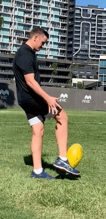
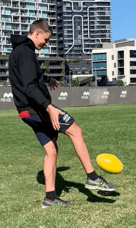
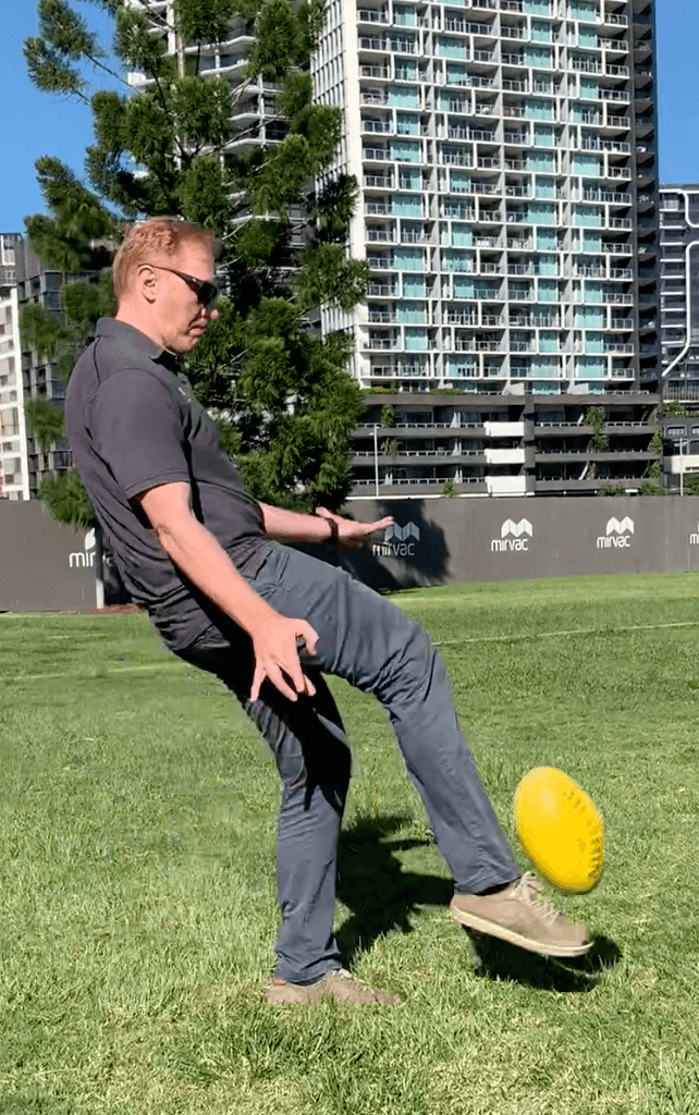
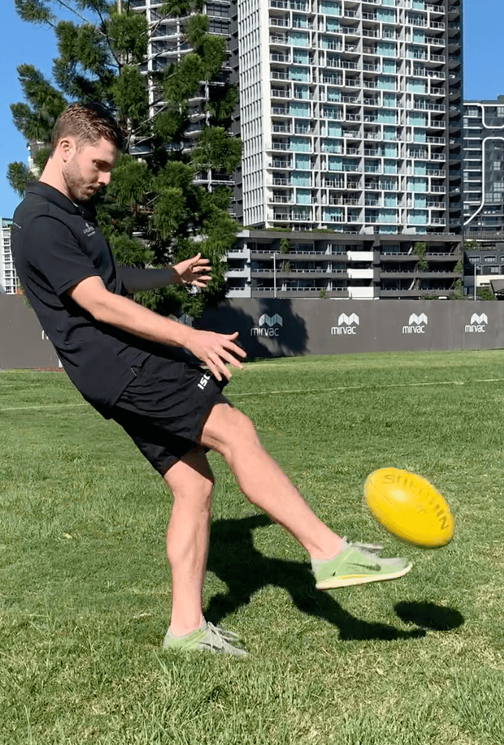
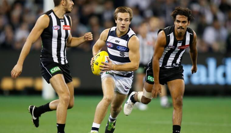
Kicking Goals! Praxis looks at AFL kicking mechanics

KICKING

As per usual the Praxis team delved into an area of clinical interest to better understand the demands of our athletes. Last session we discussed throwing technique which you can read about here.

SUMMARY

  • There are differences in kicking techniques when accuracy or distance is the focus.
  • More accurate kickers had tended to be more “head over the ball” with significantly:
  • greater hip flexion in both limbs
  • greater knee flexion in the support limb throughout the kicking movement and greater anterior pelvic tilt at heel contact
  • Longer kick distances were associated with:
  • greater foot speeds and shank angular velocities at ball contact,
  • larger last step lengths, and
  • greater distances from the ground when ball contact occurred.
  • To increase kicking distance:
  • increasing foot speed and shank angular velocity at ball contact, increasing the last step length, and
  • optimising ball position relative to the ground and support foot are recommended.
  • Injuries to the quadriceps are often associated with kicking

Given our longstanding association with AFL clubs around Brisbane, this week we looked at kicking kinematics (joint angles and relationships) with respect to both accurate and long kicks. Further, we discussed how to best rehab someone with a kicking based injury and helping them return to their chosen kicking sport (AFL, Rugby, Soccer, Gridiron etc).

ACCURACY:

The first paper we looked at was from Dichiera and colleagues (2006). Their study involved kicking an AFL football 15m at a target. They found that accurate kickers focussed more on larger hip flexion, anterior tilt and stance leg knee flexion. This best can be described as a “head over the football” approach.

They hypothesise that knee flexion is an important limb length adjustment mechanism which lowers the centre of gravity. During kicking, an increased stance leg knee bend would lower the centre of gravity somewhat throughout the movement. Lowering the centre of gravity is one way of increasing the stability of the body, a principle which is emphasised in many other areas in sports biomechanics.

DISTANCE:

Kicking for distance was associated with greater foot speeds and shank angular velocities at ball contact, larger last step lengths, and greater distances from the ground when ball contact occurred in a study performed by Ball (2008). This was more of a “lean back” strategy utilised by the kickers. Knee angular velocity at ball contact was measured at almost 1400 degrees per second! That kind of speed understandably places the knee extensors (i.e the quadriceps) at greatest risk for kicking injuries.

Ball (2008) outlined some basic coaching instructions to guide those seeking larger distances in their kicks. He suggested:

  • increasing foot speed and shank angular velocity at ball contact by increasing approach speed such that the hip of the kick leg is moving faster towards the target during the last step.
  • increasing the last step length but this step should be proportional to approach speed as over-striding is likely to be detrimental to the kick
  • Optimising ball position relative to the ground and support foot was also recommended though contact too high may result in a high kick, not a long kick.

From a physiotherapy perspective, we discussed:

  • Players have adequate hip extension range as to not to not overload lumbar spine and hip extensors (e.g hamstrings) especially in the presence of larger running and sprinting volumes typical in AFL and soccer
  • Hip flexor strengthening to improve the drive of swing leg through the kick and running
  • How to best reintegrate an athlete who sustained an injury whilst kicking back to full training and competition
  • Preventative exercise such as jump lunges and reverse nordics for quadriceps tissue resilience
  • How lumbopelvic control can help a kicker with both accuracy and distance

In summary, another Brisbane winter’s morning was successfully utilised by the Praxis team. We hope you enjoyed the read! For any injuries that you or your teammates need assessed and sorted fast, contact us.

Until next time, continue to Praxis What You Preach

– Team Praxis

​Prevent. Prepare. Perform.

References:

  1. Dichiera, A., Webster, K. E., Kuilboer, L., Morris, M. E., Bach, T. M., & Feller, J. A. (2006). Kinematic patterns associated with accuracy of the drop punt kick in Australian Football. Journal of Science and Medicine in Sport, 9(4), 292-298.
  2. Ball, K. (2008). Biomechanical considerations of distance kicking in Australian Rules football. Sports Biomechanics, 7(1), 10-23.
Throwing Injuries

Throwing Injuries

THROWING

This week in professional development session, our physio team delved into throwing techniques and links to injury. Proper throwing mechanics are important to understand as they may enable an athlete to achieve maximum performance with minimum chance of injury (Fleisig et al 2012).

Throwing, tennis serving, cricket fast bowling and golf swings are all excellent examples are how the summation of the bodies forces can result in massive outputs of power. Although force to a ball or other projectile is applied directly by the hand, a ‘kinetic chain’ of the entire body is used.

Mid Potion Achilles Tendinopathy Location

One essential and shared property of these activities is they utilise the kinetic chain to generate and transfer energy from the larger body parts to the smaller, more injury-prone upper extremities. These activities are all also notorious for high rates of injury. The kinetic chain principle asserts that in a coordinated human motion, energy and momentum are transferred through sequential body segments, achieving maximum magnitude in the terminal segment.

This kinetic chain in throwing includes the following sequence of motions: stride, pelvis rotation, upper torso rotation, elbow extension, shoulder internal rotation and wrist flexion (Fleisig et al 2012).

According to Agresta and colleagues (2019), Risk factors for shoulder pain are:

  • Workload (spikes or high volume)
  • Age (younger athletes are more prone to injury)
  • Throwing technique (e.g lack of follow through, elbow varus and shoulder external rotation torque)
  • Reduction in shoulder range of motion (particularly shoulder internal rotation in preseason)
  • Reduced preseason strength (supraspinatus and prone external rotation strength)
  • Reduction in thoracic rotation
  • Previous injury

Whilst we love a nerdy discussion on intrinsic vs extrinsic risk factors and specific rehab options as much as the next person, sometimes you just need to get outside in the sun and FEEL the task you are poring over!!

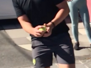
The key phases of throwing are loosely depicted in the below photos by our Praxis Principal and former 1st Grade Cricketer, Stephen (adapted from Escamilla et al 2007):

Click on the photo to slow the sequence

One of the final key questions from the day was: Who out of our physiotherapists has a “custard arm” and who has a “bullet”?? So if you or someone you know is in a throwing sport, have a chat to us today on (07) 3102 3337 or book online to ensure you have an injury free season ahead!

Until next time, Praxis what you preach

Team Praxis

Prevent. Prepare. Perform

The Single Leg Squat

The Single Leg Squat

For those of you who have ever read a research article and thought it was a tough read, i’d like to let you in on a little secret. Doing the research is far worse! If only memes were a thing when I started my Masters of Applied Science thesis, I’d have changed my background to remind myself that conducting research was even drier than every dish an apprentice has cooked for Gordon Ramsey.

My mentor at the time, who was the manager of the sports science and sports medicine devision of Cricket Australia and later the head of the AIS human movements department, Dr Marc Portus, enlightened me with something quite profound. He said, “There are two outcomes from a thesis. Either you live it for the rest of you academic days or it sits on a bookshelf for years collecting dust.” Given I completed my thesis and went straight onto my graduate entry physiotherapy masters, it is fair to say i’m in the later camp. Quite a few years have passed now however, so I’d thought i’d dust off the cover and summarise my thesis for all of you playing at home!

So not to degrade my self entirely, I thought I found some pretty good stuff that has affected the way I assess and treat today. I haven’t shared it all today as it was more than 140 pages long and ‘ain’t nobody got time for that’. More importantly though, my thesis reminded me of the passion I have for lower limb biomechanics and that physiotherapy (as opposed to research) was always meant to be on the cards for me. My thesis, “The 3D Kinematics of the Single Leg Flat and Decline Squats” boiled down to looking at how the ankle position changed the joint angles single leg squat, how hip strength affected the squat as well as few other things that aren’t worth mentioning here.

BACKGROUND:

The single leg squat (SLS) replicates an athletic position commonly assumed in sport such as cutting (powerful change in direction while running made from one leg), jumping and balancing which all require the control of the trunk and pelvis on the weight bearing femur in all three planes of movement [1-5].

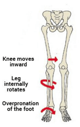
As such, the SLS is commonly used by clinicians as a functional measure of dynamic lumbo-pelvic stability [endif]–[6-8]. Abnormal movement within the SLS tend to be characterised by the commonly described “medial collapse” or “dynamic valgus”. Specifically, there is excessive femoral internal rotation, femoral adduction, knee valgus, tibial internal rotation and foot pronation of the weight-bearing limb with resultant excursion of the contralateral non weight bearing Ilium and excessive lateral flexion of the trunk [endif]–[3, 6-8].

The reason why this tends to be perceived as a big deal is that this position tends to be argued as a lack of lumbopelvic stability and results in increased loading of the knee. Moreover, pelvis weakness tends to be ascribed to the absence of stability ultimately resulting in a position in which many acute and overuse injuries of the lower limb may occur. These ailments include, ACL / MCL ruptures, patellofemoral pain syndrome (PFPS), illiotibial band friction syndrome (ITBFS) and shin splints to name a few. That is why the SLS appears to be a valuable rough screening tool in clinical practice.

MY FINDINGS:

As mentioned, I looked at how a decline board of 20 degrees changed the angles of the lower limb during the squat. I also looked at if any strength measures of the hip related to how someone squatted between conditions. Finally, I looked at if the decline board altered how someone was scored by experienced physiotherapist as a competent or not at the squat

JOINT ANGLES (KINETMATICS:)

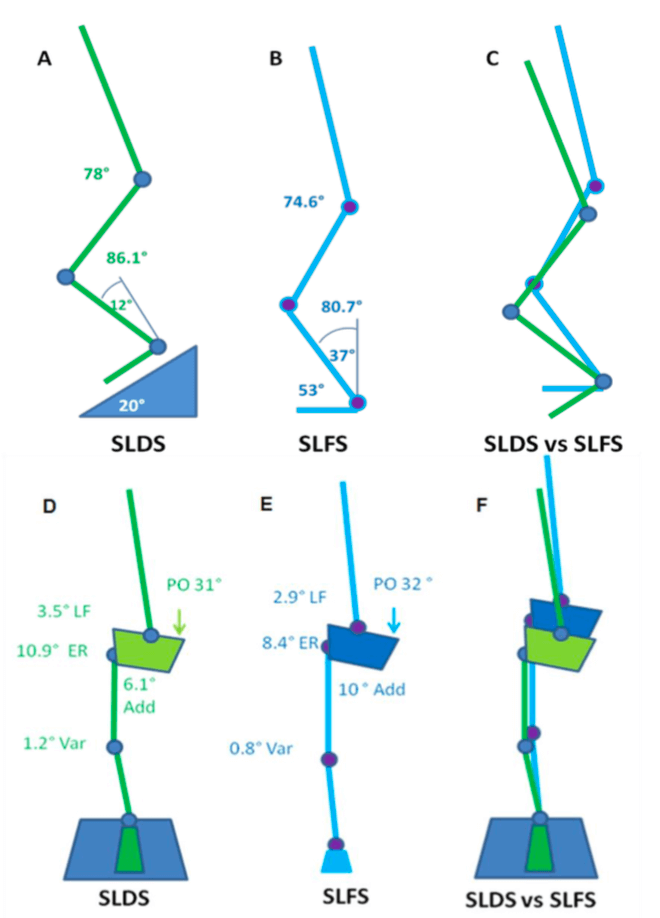
A picture tells a thousand words so in the interests of brevity, the stick squat figure is essentially a summary of two years of work.

So what this means, when someone performs a SLS on a flat surface, relative to a decline surface they tend to have:

  • A more upright torso
  • More rotation of the pelvis toward the weigh bearing (WB) limb
  • Reduced flexion but more adduction and internal rotation of the thigh on the WB hip (pelvic close to femur)
  • Less flexion of the knee but the same position relative to the foot as you look from the front (known as frontal plane knee excursion) at the bottom of range
  • Reduced internal rotation of the shin
  • Reduced ankle flexion

Essentially, in a flat squat you tend to ‘corkscrew’ your pelvis and adopt the medial collapse position much more easily than in the decline squat position. This may because of ankle range of motion issues as well as the ability to adequately recruit pelvic musculature. Yep – two years to get that!

STRENGTH AND MOVEMENT:

My results demonstrated a tendency for the pelvis to remain increasingly level with greater hip abduction strength. However, the relationship between strength and the pelvis was observed in the decline condition but not the flat condition. This may be due to hip abduction was shown to be significantly less (more neutral) in the SLDS which seemingly promoted greater muscle activation and subsequent control of pelvis. The self selection of squat depth may have also been a critical factor in finding as those with weak hips may have squatted deep to adopt maladaptive positions. Previous research has indicated that the hip abductors and external rotators play an important role in lower extremity alignment as they assist in the maintenance of a level pelvis [9] and are capable in balancing a number of biomechanical forces in the body [10].

Interestingly, there were no significant relationships observed between hip abduction strength and knee valgus (knee falling in) for both squatting conditions. There was however a trend between hip abduction strength and knee valgus which supported previous research. It is keeping with the assumption that increased knee valgus might also be associated with reduced hip abduction and external rotation strength [11].

SUMMARY:

  • To maximise athletic function, particularly in sports such as soccer, netball and AFL, stability through the pelvis and hips, proximal lower limb, spine and abdominal structures is required [12].
  • The importance of pelvis stabilisation for lower extremity injury prevention [13] particularly the knee [14-17] has been well documented in the literature.
  • Adequate lumbopelvic-femur strength and muscle function may conceivably reduce exposure to other intrinsic risk factors such as inefficient force attenuation, unstable movement patterns and lower limb malalignments during activity [18, 19].
  • Ankle flexibility may also be a factor in lower limb physical resilience and injury prevention.
  • Support for the previous statements has been demonstrated in the relationships between hip strength measures and kinematics within selected results of my study.

There you have it. Two years of my life summarised to a few paragraphs. From a personal perspective, I took away from my research experience to be always questioning why we do things and see if there is someone out there who has answered the questions we seek. Finally, don’t overcook chicken – Ramsay doesn’t like it.
REFERENCES:

  1. Neely, F.G., Intrinsic risk factors for exercise-related lower limb injuries. Journal of Sports Medicine, 1998. 26(4): p. 253-263.
  2. Parkkari, J., U.M. Kujala, and K. Pekka, Is it possible to prevent sports injuries? Review of controlled clinical trials and recommendations for future work. Sports Medicine, 2001. 31(14): p. 985-995.
  3. Lysens, R.J., et al., The accident -prone and overuse-prone profiles of the young athlete. The American Journal of Sports Medicine, 1989. 17(5): p. 612-619.
  4. Egger, G., Sports injuries in Australia: causes, costs and prevention. A report to the national better health program., ed. C.f.H.P.a. Research. 1990, Sydney.
  5. Orchard, J.W. and C.F. Finch, Australia needs to follow New Zealand’s lead on sports injuries. The Medical Journal of Australia, 2002. 177: p. 38-39.
  6. Wu, G. and P.R. Cavanagh, ISB recommendations for standardization in the reporting of kinematic data. Journal of Biomechanics, 1995. 28: p. 1257- 1261.
  7. Siegal, P., R. Brackbill, and G. Heath, The epidemiology of walking exercise: implications for promoting activity among sedentary groups. American Journal of Public Health, 1995. 85(5): p. 706-710.
  8. Nicholl, J.P., P. Coleman, and B.T. Williams, The epidemiology of sports and exercise related injury in the United Kingdom. British Journal of Sports Medicine, 1995. 29(4): p. 232-238.
  9. Burnet, E.N. and P.E. Pidcoe, Isometric gluteus medius muscle torque and frontal plane pelvic motion during running. Journal of Sports Science and Medicine, 2009. 8: p. 284-288
  10. Niemuth, P., et al., Hip muscle weakness and overuse injuries in recreational runners. Clinical Journal of Sports Medicine, 2005. 15(1): p. 14-21.
  11. Hollman, J.H., et al., Relationships between knee valgus, hip-muscle strength, and hip-muscle recruitment during a single-limb step down. Journal of Sport Rehabilitation, 2009. 18: p. 104-117.
  12. Kibler, W.B., J. Press, and A. Sciascia, The role of core stability in the athletic function Journal of Sports Medicine, 2006. 36(3): p. 189-198.
  13. Leetun, D.T., et al., Core stability measures as risk factors for lower extremity injury in athletes. Medicine & Science in Sports & Exercise, 2004. 36(6): p. 926-934.
  14. Cichanowski, H., et al., Hip strength in collegiate female athletes with patellofemoral pain. Medicine & Science in Sport & Exercise, 2007. 39(8): p. 1227-1232.
  15. Ireland, M.L., et al., Hip strength measures in female with and without patellofemoral pain. Journal of Orthopaedic & Sports Physical Therapy, 2003. 33(11): p. 671-676.
  16. Nicholas, J.A., A.M. Strizak, and G. Veras, A study of thigh muscle weakness in different pathological states of the lower extremity. American Journal of Sports Medicine, 1976. 4: p. 241-248.
  17. Prins, M.R. and P.V.D. Wurff, Females with patellofemoral pain syndrome have weak hip muscles: a systematic review. Australian Journal of Physiotherapy, 2009. 55: p. 9-15.
  18. Willson, J.D., M.L. Ireland, and I. Davis, Core strength and lower extremity alignment during single leg squats. Medicine & Science in Sports & Exercise, 2006. 38(5): p. 945-952.
  19. Lee, D., The pelvic girdle: An approach to the examination and treatment of the lumbopelvic-hip region. 3rd ed. 2004, Edinburugh: Churchill Livingston.
Timms on Tour (again)

Timms on Tour (again)

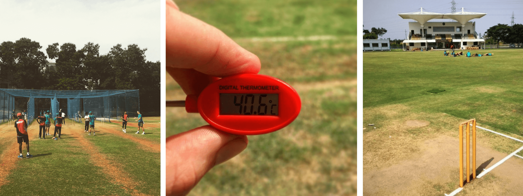
Another month and another Cricket Australia tour it seems for our Praxis Principal, Stephen Timms. Stephen is providing strength, conditioning and physiotherapy support at the training camp to the MRF Academy in Chennai as a part of the National Performance Squad (NPS).

The NPS program was formerly known as the Australian Cricket Academy and the Centre of Excellence. The program is designed to add to the work done within professional state systems, and prepare players capable of competing at international level for Australia within the next five years.

The MRF Academy tour has a strong focus on spin and adapting to subcontinental conditions, which can include oppressive heat and humidity. The NPS will be mentored by former Test players Chris Rogers and Ryan Harris along with NPS head coach Troy Cooley.

The squad typically spends more than three months at the Brisbane based Bupa National Cricket Centre. Here they have access to world class facilities and coaching and utilise the typically dry and sunny Brisbane winters to make the most of the off season program.

Recent graduates of the program who have gone on to play for Australia include Alex Carey, Matthew Renshaw and Hilton Cartwright.

Stephen has arrived back in Australia but will continue to service the NPS until the end of a series of interstate games September. He will be back in the clinic around mid September. If you are looking for an appointment before that time, please call our central number (07) 3102 3337 so we can book you in with one of our other skilled therapists.

We are always excited to see Stephen and all our therapists flying the Praxis flag and applying the Prevent Prepare Perform motto in elite sport!

For more details on the program and players selected in the NPS, check out the link to the Cricket Australia website here.

If you are Cricketer and looking to Prevent Perpare Perform, book in online with Stephen or one of our other skilled therapists at Praxis Physiotherapy.

Yours in Health,

The Praxis Team

Prevent. Prepare. Perform.