Knee Osteoarthritis: Is ‘Bone on Bone’ a painful life sentence?

Knee Osteoarthritis: Is ‘Bone on Bone’ a painful life sentence?

  • Knee Osteoarthritis is a common ailment responsible for pain, loss of function and reduced quality of life
  • Rates of knee OA are set to increase
  • Whilst there is no cure, exercise therapy under the guidance of a physiotherapist is considered a front line treatment to help reduce the severity of symptoms
  • There are options before a knee replacement

Do your knees go crackle and pop? Pain with walking, stairs or getting out of a chair? Stiffness and pain first thing in the morning or after a long car ride? These are signs that you may be living with the early or even advanced symptoms of knee osteoarthritis (OA). Don’t fear though – there is plenty that can be done immediately.

What is “OA”?

Osteoarthritis (OA) is an increasingly prevalent source of musculoskeletal pain and dysfunction. OA is a disease of the joint – including cartilage, bone, capsule and other associated tissues. This disease process can cause chronic pain, reduced physical function and diminished quality of life. The ageing population and increased global prevalence of obesity are anticipated to dramatically increase the impacts of knee OA and its associated impairments [1]. Although osteoarthritis can affect any joint, OA is knee is one of the most common complaints.

Presentation

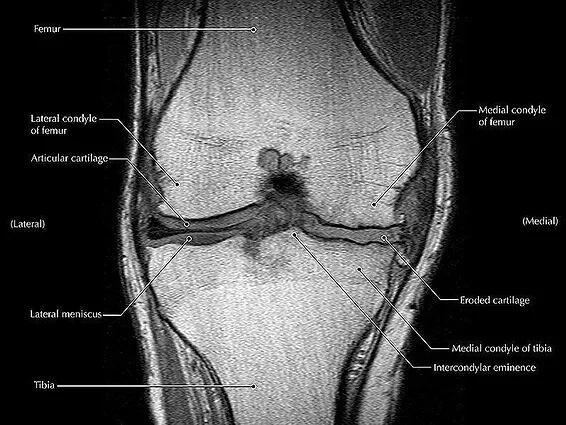
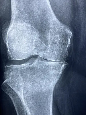
It most commonly presents in people over the age of 50, and is often described as being painful, stiff and occasionally swollen. In terms of a tissue level, knee OA describes the gradual deterioration of the supportive cartilage within the knee joint. As the cartilage wears away with time, the protective joint space between the bones decreases. With a reduced cartilage lining to protect and support the spacing of the knee joint, the Femur and Tibia (knee bones) are increasingly less likely to dissipate forces through the joint . With time, it should be expected that bone spurs (osteophytes) may form in and around the joint as the bones react to repetitive contact with each other.

Management

The management of knee OA largely consists of exercises addressing strength, range of motion, quality of movement, emphasizing joint control, pain reduction and weight management.

Strength Training

Strength training should be the cornerstone of addressing knee OA, particularly the early signs. Strengthening the muscles around the knee joint, such as the quadriceps, hamstrings, and glutes provide better support to the knee, reducing stress on the joint and helping to alleviate pain and discomfort. Movement associated with exercise has an added benefit – It increases joint lubrication. Loading of the joint stimulates the production and distribution of synovial fluid within the joint. This fluid acts as a lubricant, reducing friction and providing cushioning to the joint surfaces. Improved lubrication can help alleviate pain during movement.

Range of motion

Knee osteoarthritis often leads to stiffness and limited range of motion in the joint. Physiotherapy can include specific exercises, manual therapy and stretches to improve joint flexibility, helping to restore a more normal range of motion and enhancing mobility. The greater the restoration of range, the better the knee feels.

Pain reduction

Both strength training and physiotherapy can help reduce pain associated with knee OA. As mentioned, stronger muscles provide better support to the joint, relieving pressure and reducing pain during movement. Physiotherapy may provide education of aggravating and easing factors (eg. hot / cold packs, hydrotherapy) as well as liaise with your GP for adequate analgesic medications.

Lifestyle modifications

Adopting a healthy lifestyle can play a pivotal role in managing knee osteoarthritis. Maintaining a healthy weight reduces the stress on the knee joints. Regular low-impact exercises such as swimming, cycling and reformer pilates help improve strength, flexibility, and overall joint health. A balanced diet rich in fruits, vegetables, whole grains, and lean proteins can promote weight loss and provide essential nutrients for joint health. Quitting smoking and minimizing alcohol consumption are also beneficial.

Improved weight management

Regular exercise can assist in weight management, which is crucial for individuals with knee osteoarthritis. Excess weight puts additional strain on the knee joint, contributing to pain and progression of the condition. By maintaining a healthy weight, exercise helps to reduce the load on the joint and alleviate pain.

Surgical Interventions

When conservative measures fail to provide relief, surgical interventions may be necessary. Procedures such as arthroscopy, osteotomy, and joint replacement surgery can help repair damaged tissues, realign the joint, or replace the damaged joint with a prosthetic. These surgeries can significantly improve mobility and reduce pain, allowing individuals to resume their daily activities. Physiotherapy can aid in preparing you for the surgery, as well as rebuild your “new” knee after a knee replacement has been completed.

In conclusion, while knee osteoarthritis can be challenging, it is not a condition that should hinder individuals from leading fulfilling lives. By implementing lifestyle modifications, exploring various treatment options, and working closely with your physiotherapist, individuals can effectively manage their symptoms, alleviate pain, and enjoy an active lifestyle with a sense of well-being. If conservative options fail, there are surgical interventions that can be investigated. If you are wanting to look after your knees, or already suffering from knee pain, chat to our knowledgeable Praxis Physios to discuss your treatment options at any stage of OA’s progression.

Until next time,

Praxis what you Preach

Jumper’s Knee (Patellar Tendinopathy)

Jumper’s Knee (Patellar Tendinopathy)

Do you play a jumping sport such as volleyball, basketball or AFL? Have pain in the front of your knee when jumping, landing or changing direction? Have you lost some jumping power recently? Well read on friends as you may have a grumbly knee tendon.

Summary:

  • Patellar tendinopathy is summarised clinically as pain and dysfunction in the patellar tendon
  • Most commonly affects jumping athletes from adolescence to early middle age.
  • Return to sport can be slow with physio useful as a front line management tool
  • Often requires prolonged rehabilitation centred around education, strength training and load management

Jumper’s knee (or patellar tendinopathy) as its name suggest predominantly affects athletes who engage in sports which require large volumes of jumping. Jumping dynamically loads the knee and places large loads on the patellar tendons due the large and repeated requirements of the thigh muscles (quadriceps). These include sports that require repeat jump / landing efforts and/or high volumes of load during training and competition. Elite adolescent male athletes tend to be at a higher risk, especially if you play volleyball.

Mid Potion Achilles Tendinopathy Location

Like most injuries, patellar tendinopathy reflects an overload of the tissue and a failed healing response. Tendons tend to most susceptible to long periods of dynamic loading given their role in storing and releasing energy like a spring. The stiffer the spring, the more effective the spring and the more punishment it can take before the function deteriorates.

This injury is one that can be mild or moderate in nature and as such allow playing to some degree. As such, player’s tend to not to miss a lot of games like more “traditional” injuries such as ankle sprains or hamstring tears. It can typically slowly present and have a “warm up phenomena” (as in it can get better during a game), however aches after activity and the next morning. The pain is often at the very bottom of the knee cap, and on the space between the kneecap and the top of the shin bone where the tendon lies.

Key management strategies include ensuring the correct diagnosis and an understanding of tendon pathology (for more on tendon pathology, check out this blog). From there, pain management strategies and workload management is a key tenant to rehabilitation. Above and beyond workload management and good patient education, we at Praxis Physio also test the strength and range of the hip, knee and ankle musculature as well as jumping / landing biomechanics to understand where the likely reasons are for your knee pain.

After a comprehensive assessment, targeted and graduated strengthening is provided. The premise of these early phases are to reduce pain, improve strength, improve function, increase power (specifically the energy storage potential of the tendon) then finally sports specific training and management on symptoms.

As someone who has had an 18 month history of patellar tendinopathy, I personally can attest to the frustration this injury provides. I made many mistakes along my rehabilitation journey – though this was before I was a physiotherapist and took a clinical interest in tendinopathies. Thankfully, the research has come a long way in the last decade, so if you are having ongoing knee pain that you suspect is jumper’s knee, book in with us so we can get you jumping back to your best.

Until next time, Praxis what you Preach.

Stephen Timms

Knee Pain

Knee Pain

Do you have a good knee and a bad knee? Do your knees snap, crackle and pop? Do you ever wish you could walk for longer without pain or not be worried about how your knees will feel in the morning if you were living your life to the fullest? Knee pain is a very common condition that affects people of all ages. Typically knee pain is the result of an injury on the sporting field when we are younger but tend to be more degenerative in nature as we age.
Mid Potion Achilles Tendinopathy Location

Different knee injuries tend to have differing symptoms. Common symptoms include:

  • Aching, sharp, stabbing and/or catching pains
  • Large amounts of swelling or sometimes pockets of swelling
  • Warm to touch
  • Feelings of grating, grinding or even giving way
  • Pops and crunching noises
  • Unable to full bend or straighten the knee

The structures of the knee that are often implicated in knee pain are the patellar or quadriceps tendons, cartilage, meniscus, bursas, and even major ligaments such as the anterior cruciate ligament, otherwise known as the ACL.

Common injuries to the knee can be:

  • Osteoarthritis
  • Tendinopathy / Tendonitis
  • Bursitis
  • Knee cap pain (patellofemoral pain)
  • Meniscal tear (degenerative and acute tears)
  • Dislocated knee cap
  • Iliotibial band friction syndrome

Regardless of your symptoms and presentation, our highly trained Praxis physiotherapists have expertise in this area and will help identify the problem and work with you so that you will feel empowered to fix the problem. At Praxis Physio this is our point of difference, we promise to take the time to fix you using a range of modalities including advice, hands on manual therapies and of course strength and conditioning programming. In addition, we work in close collaboration with leading knee surgeons if this course of action is required.


There is no need to accept knee pain as ‘normal’. Call us now on (07) 3102 3337 or book online to have one of our physios develop a plan to reduce your pain and restore your function!

To read more about how running can help your knees (that’s right – running!) check out our related posts on running written by our published principal physio, Stephen.

Team Praxis,

PREVENT | PREPARE | PERFORM

FACT OR FICTION FRIDAY || Knee Pain and Scans

FACT OR FICTION FRIDAY || Knee Pain and Scans

Answer: FICTION 🙊

“Osteoarthritis” is a common term that gets used by our patients as an explanation of their knee pain. But is this always the case 🤔?

🔍In a recent systematic review estimates of osteoarthritis feature prevalence on MRI among asymptomatic uninjured knees were up to 14% in adults < 40 years, and up to 43% in adults > 40 years!

Whilst features on MRI imaging such as cartilage defects, meniscal tears and osteophyte lesions can potentially play a role if you have pain, this should always be interpreted in the context of your clinical presentation by a health care professional as these changes can be normal in an asymptomatic population – just like grey hair as we age 👴👵!

If you have knee pain and have resigned yourself to a ‘life sentence’, come and have a chat to one of our physios to ensure you aren’t robbing yourself of a full functioning future 🕺🏃🏌️🏄🏋️🏊🚴🏂🎾! Call 07 3102 3337 or book online 

#kneeoa #praxisphysio #factorfictionfriday #preventprepareperform #kneepain #kneeosteoarthritis #mri #fullfunctionfuture

Reference:

Culvenor AG, Øiestad BE, Hart HF, et al Prevalence of knee osteoarthritis features on magnetic resonance imaging in asymptomatic uninjured adults: a systematic review and meta-analysis British Journal of Sports Medicine 2019;53:1268-1278.

The Single Leg Squat

The Single Leg Squat

For those of you who have ever read a research article and thought it was a tough read, i’d like to let you in on a little secret. Doing the research is far worse! If only memes were a thing when I started my Masters of Applied Science thesis, I’d have changed my background to remind myself that conducting research was even drier than every dish an apprentice has cooked for Gordon Ramsey.

My mentor at the time, who was the manager of the sports science and sports medicine devision of Cricket Australia and later the head of the AIS human movements department, Dr Marc Portus, enlightened me with something quite profound. He said, “There are two outcomes from a thesis. Either you live it for the rest of you academic days or it sits on a bookshelf for years collecting dust.” Given I completed my thesis and went straight onto my graduate entry physiotherapy masters, it is fair to say i’m in the later camp. Quite a few years have passed now however, so I’d thought i’d dust off the cover and summarise my thesis for all of you playing at home!

So not to degrade my self entirely, I thought I found some pretty good stuff that has affected the way I assess and treat today. I haven’t shared it all today as it was more than 140 pages long and ‘ain’t nobody got time for that’. More importantly though, my thesis reminded me of the passion I have for lower limb biomechanics and that physiotherapy (as opposed to research) was always meant to be on the cards for me. My thesis, “The 3D Kinematics of the Single Leg Flat and Decline Squats” boiled down to looking at how the ankle position changed the joint angles single leg squat, how hip strength affected the squat as well as few other things that aren’t worth mentioning here.

BACKGROUND:

The single leg squat (SLS) replicates an athletic position commonly assumed in sport such as cutting (powerful change in direction while running made from one leg), jumping and balancing which all require the control of the trunk and pelvis on the weight bearing femur in all three planes of movement [1-5].

As such, the SLS is commonly used by clinicians as a functional measure of dynamic lumbo-pelvic stability [endif]–[6-8]. Abnormal movement within the SLS tend to be characterised by the commonly described “medial collapse” or “dynamic valgus”. Specifically, there is excessive femoral internal rotation, femoral adduction, knee valgus, tibial internal rotation and foot pronation of the weight-bearing limb with resultant excursion of the contralateral non weight bearing Ilium and excessive lateral flexion of the trunk [endif]–[3, 6-8].

The reason why this tends to be perceived as a big deal is that this position tends to be argued as a lack of lumbopelvic stability and results in increased loading of the knee. Moreover, pelvis weakness tends to be ascribed to the absence of stability ultimately resulting in a position in which many acute and overuse injuries of the lower limb may occur. These ailments include, ACL / MCL ruptures, patellofemoral pain syndrome (PFPS), illiotibial band friction syndrome (ITBFS) and shin splints to name a few. That is why the SLS appears to be a valuable rough screening tool in clinical practice.

MY FINDINGS:

As mentioned, I looked at how a decline board of 20 degrees changed the angles of the lower limb during the squat. I also looked at if any strength measures of the hip related to how someone squatted between conditions. Finally, I looked at if the decline board altered how someone was scored by experienced physiotherapist as a competent or not at the squat

JOINT ANGLES (KINETMATICS:)

A picture tells a thousand words so in the interests of brevity, the stick squat figure is essentially a summary of two years of work.

So what this means, when someone performs a SLS on a flat surface, relative to a decline surface they tend to have:

  • A more upright torso
  • More rotation of the pelvis toward the weigh bearing (WB) limb
  • Reduced flexion but more adduction and internal rotation of the thigh on the WB hip (pelvic close to femur)
  • Less flexion of the knee but the same position relative to the foot as you look from the front (known as frontal plane knee excursion) at the bottom of range
  • Reduced internal rotation of the shin
  • Reduced ankle flexion

Essentially, in a flat squat you tend to ‘corkscrew’ your pelvis and adopt the medial collapse position much more easily than in the decline squat position. This may because of ankle range of motion issues as well as the ability to adequately recruit pelvic musculature. Yep – two years to get that!

STRENGTH AND MOVEMENT:

My results demonstrated a tendency for the pelvis to remain increasingly level with greater hip abduction strength. However, the relationship between strength and the pelvis was observed in the decline condition but not the flat condition. This may be due to hip abduction was shown to be significantly less (more neutral) in the SLDS which seemingly promoted greater muscle activation and subsequent control of pelvis. The self selection of squat depth may have also been a critical factor in finding as those with weak hips may have squatted deep to adopt maladaptive positions. Previous research has indicated that the hip abductors and external rotators play an important role in lower extremity alignment as they assist in the maintenance of a level pelvis [9] and are capable in balancing a number of biomechanical forces in the body [10].

Interestingly, there were no significant relationships observed between hip abduction strength and knee valgus (knee falling in) for both squatting conditions. There was however a trend between hip abduction strength and knee valgus which supported previous research. It is keeping with the assumption that increased knee valgus might also be associated with reduced hip abduction and external rotation strength [11].

SUMMARY:

  • To maximise athletic function, particularly in sports such as soccer, netball and AFL, stability through the pelvis and hips, proximal lower limb, spine and abdominal structures is required [12].
  • The importance of pelvis stabilisation for lower extremity injury prevention [13] particularly the knee [14-17] has been well documented in the literature.
  • Adequate lumbopelvic-femur strength and muscle function may conceivably reduce exposure to other intrinsic risk factors such as inefficient force attenuation, unstable movement patterns and lower limb malalignments during activity [18, 19].
  • Ankle flexibility may also be a factor in lower limb physical resilience and injury prevention.
  • Support for the previous statements has been demonstrated in the relationships between hip strength measures and kinematics within selected results of my study.

There you have it. Two years of my life summarised to a few paragraphs. From a personal perspective, I took away from my research experience to be always questioning why we do things and see if there is someone out there who has answered the questions we seek. Finally, don’t overcook chicken – Ramsay doesn’t like it.

REFERENCES:

  1. Neely, F.G., Intrinsic risk factors for exercise-related lower limb injuries. Journal of Sports Medicine, 1998. 26(4): p. 253-263.
  2. Parkkari, J., U.M. Kujala, and K. Pekka, Is it possible to prevent sports injuries? Review of controlled clinical trials and recommendations for future work. Sports Medicine, 2001. 31(14): p. 985-995.
  3. Lysens, R.J., et al., The accident -prone and overuse-prone profiles of the young athlete. The American Journal of Sports Medicine, 1989. 17(5): p. 612-619.
  4. Egger, G., Sports injuries in Australia: causes, costs and prevention. A report to the national better health program., ed. C.f.H.P.a. Research. 1990, Sydney.
  5. Orchard, J.W. and C.F. Finch, Australia needs to follow New Zealand’s lead on sports injuries. The Medical Journal of Australia, 2002. 177: p. 38-39.
  6. Wu, G. and P.R. Cavanagh, ISB recommendations for standardization in the reporting of kinematic data. Journal of Biomechanics, 1995. 28: p. 1257- 1261.
  7. Siegal, P., R. Brackbill, and G. Heath, The epidemiology of walking exercise: implications for promoting activity among sedentary groups. American Journal of Public Health, 1995. 85(5): p. 706-710.
  8. Nicholl, J.P., P. Coleman, and B.T. Williams, The epidemiology of sports and exercise related injury in the United Kingdom. British Journal of Sports Medicine, 1995. 29(4): p. 232-238.
  9. Burnet, E.N. and P.E. Pidcoe, Isometric gluteus medius muscle torque and frontal plane pelvic motion during running. Journal of Sports Science and Medicine, 2009. 8: p. 284-288
  10. Niemuth, P., et al., Hip muscle weakness and overuse injuries in recreational runners. Clinical Journal of Sports Medicine, 2005. 15(1): p. 14-21.
  11. Hollman, J.H., et al., Relationships between knee valgus, hip-muscle strength, and hip-muscle recruitment during a single-limb step down. Journal of Sport Rehabilitation, 2009. 18: p. 104-117.
  12. Kibler, W.B., J. Press, and A. Sciascia, The role of core stability in the athletic function Journal of Sports Medicine, 2006. 36(3): p. 189-198.
  13. Leetun, D.T., et al., Core stability measures as risk factors for lower extremity injury in athletes. Medicine & Science in Sports & Exercise, 2004. 36(6): p. 926-934.
  14. Cichanowski, H., et al., Hip strength in collegiate female athletes with patellofemoral pain. Medicine & Science in Sport & Exercise, 2007. 39(8): p. 1227-1232.
  15. Ireland, M.L., et al., Hip strength measures in female with and without patellofemoral pain. Journal of Orthopaedic & Sports Physical Therapy, 2003. 33(11): p. 671-676.
  16. Nicholas, J.A., A.M. Strizak, and G. Veras, A study of thigh muscle weakness in different pathological states of the lower extremity. American Journal of Sports Medicine, 1976. 4: p. 241-248.
  17. Prins, M.R. and P.V.D. Wurff, Females with patellofemoral pain syndrome have weak hip muscles: a systematic review. Australian Journal of Physiotherapy, 2009. 55: p. 9-15.
  18. Willson, J.D., M.L. Ireland, and I. Davis, Core strength and lower extremity alignment during single leg squats. Medicine & Science in Sports & Exercise, 2006. 38(5): p. 945-952.
  19. Lee, D., The pelvic girdle: An approach to the examination and treatment of the lumbopelvic-hip region. 3rd ed. 2004, Edinburugh: Churchill Livingston.