Strong Bones, Strong Runner: Understanding and Treating Stress Fractures

Strong Bones, Strong Runner: Understanding and Treating Stress Fractures

Understanding Stress Fractures in Runners: Risk, Recovery, and Prevention

Stress fractures are a frustrating reality for many runners. Characterised by small cracks or severe bone reactions due to repetitive load, these injuries can derail training for weeks or months, and in some cases, end seasons or careers. While they are most commonly associated with endurance sports like distance running, the underlying mechanisms are multifactorial and complex. This blog explores the current understanding of stress fractures in runners — including emerging research, rehabilitation strategies, and how to lower your injury risk.

What Is a Stress Fracture?

A stress fracture is a type of bone stress injury (BSI), an overuse injury caused by the accumulation of microdamage in bone tissue due to repeated loading. Unlike acute fractures that result from a single traumatic event, stress fractures occur when repetitive sub-threshold forces — like running — outpace the bone’s capacity to repair itself (Hoenig et al., 2022).

Bone is a dynamic tissue that remodels in response to stress. However, when this remodeling process cannot keep up with microdamage accumulation — due to either an increase in training load or inadequate recovery — bone strength deteriorates. This can progress from a stress reaction to a stress fracture and, if untreated, to a complete fracture (Bergman & Kaiser, 2025; Coslick et al., 2024).

Why Are Runners So Prone?

Running, by nature, imposes repeated high loads on the lower limbs. The tibia (shin bone), metatarsals, femur, and pelvis are frequent stress fracture sites in runners (Hadjispyrou et al., 2023). Several factors contribute to the elevated risk in this group:

  • Training Errors: Rapid increases in volume or intensity, excessive hill work, or high mileage without adequate rest periods.

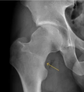
  • Bone Geometry: Martin & Heiderscheit (2023) found associations between proximal femur geometry and increased stress fracture risk, suggesting that individual anatomical differences can affect how load is distributed through the skeleton.

  • Energy Deficiency: Low energy availability, often associated with disordered eating or high training demands, can impair bone remodeling and increase injury risk — particularly in female athletes.

  • Surface and Footwear: Hard surfaces, old or inappropriate shoes, and poor running biomechanics can all contribute to abnormal load distribution and localized bone stress.

High-Risk vs Low-Risk Locations

Not all stress fractures are created equal. According to Coslick et al. (2024), stress fractures are categorized based on location and associated risk of complications:

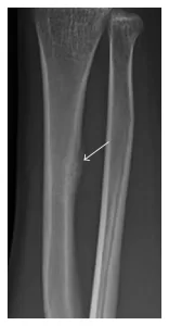
  • Low-risk sites (e.g., posterior tibia, fibula, second metatarsal shaft) typically heal well with conservative treatment.

  • High-risk sites (e.g., anterior tibia, navicular, femoral neck, and sacrum) are more likely to progress to non-union or full fracture and may require surgical management.

A nuanced understanding of the fracture location helps guide both treatment duration and rehabilitation intensity.

The Cumulative Risk Concept

Traditional models have viewed stress fractures as the result of isolated risk factors. However, Hamstra-Wright et al. (2021) propose a more integrated concept: the cumulative risk profile. This model acknowledges that risk factors — like energy deficiency, training load spikes, biomechanics, menstrual history, and previous BSIs — rarely occur in isolation.

In this framework, stress fractures occur when the athlete’s “load capacity” is exceeded by their “training load.” What’s striking is that two runners could follow the same training program but respond very differently based on their individual capacity, bone density, and recovery habits.

Clinically, this means runners must be assessed holistically. It also underscores the importance of individualized training plans, particularly during return-to-run phases.

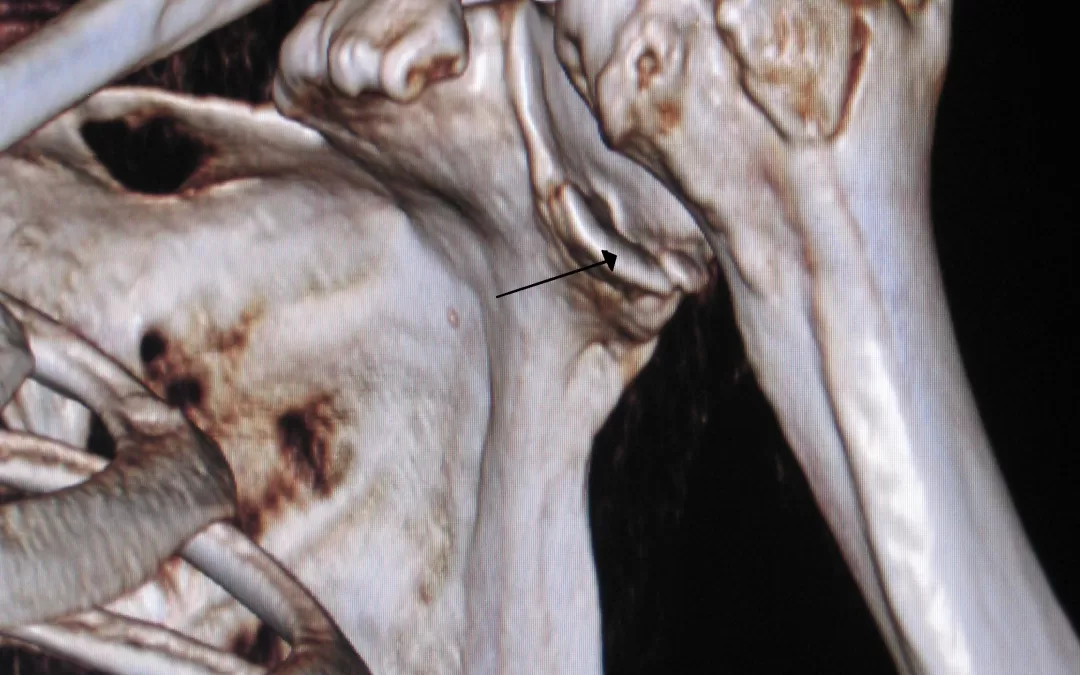
Diagnosis and Imaging

Early symptoms of a stress fracture include localized pain that worsens with activity and settles with rest. As the injury progresses, pain can persist with walking or even at rest.

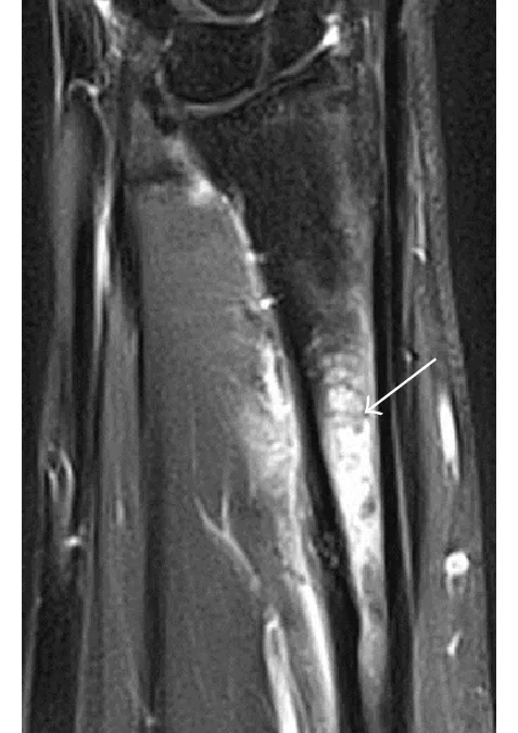
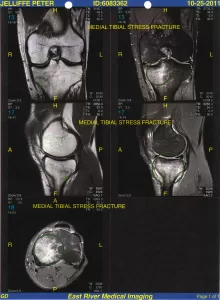
Unfortunately, standard X-rays often miss early bone stress injuries. MRI is the gold standard, able to detect bone marrow edema (early stress reaction) before a fracture line develops (Coslick et al., 2024; Bergman & Kaiser, 2025). Bone scans and CT can also be used in specific cases.

Rehabilitation and Return to Running

The cornerstone of stress fracture management is load reduction, typically involving rest from impact activities for 4–8 weeks depending on the site and severity. During this time, runners can usually continue cross-training (e.g., cycling, swimming) to maintain cardiovascular fitness.

A gradual return-to-run program should be guided by symptom response, starting with walk–run intervals and progressing to continuous running. Strength and conditioning plays a vital role in both rehabilitation and prevention — building muscular resilience to offload bony structures. Calf, hip, and core-focused strength work can significantly reduce recurrence risk and should form part of a comprehensive return-to-run strategy. (You can learn more about how we use strength and conditioning at Praxis Physiotherapy to support our runners here)

Coslick et al. (2024) emphasises the value of a multidisciplinary approach involving physiotherapists, sports physicians, dietitians, and coaches.

Preventing Stress Fractures: What Runners Can Do

While not all BSIs are preventable, runners can reduce their risk by addressing modifiable factors:

  • Progress training gradually: Avoid spikes in weekly mileage (>10% per week) and ensure at least one rest day.

  • Fuel adequately: Runners with low energy availability are at significantly increased risk, particularly females with menstrual disturbances.

  • Build strength: Muscle fatigue reduces shock absorption. Strengthening the calves, glutes, and trunk can reduce bone loading.

  • Check your shoes and form: Replace runners every 500–800 km and consider a running gait assessment, especially if you have a history of injury.

  • Listen to your body: Early symptoms like persistent aching, pinpoint bony pain, or pain that lingers after a run shouldn’t be ignored.

The Bottom Line

Stress fractures in runners are complex, multifactorial injuries that require a careful balance of training load, nutrition, and recovery. While new imaging and biomechanics research has enhanced our ability to diagnose and understand them, the best approach remains holistic — considering both the runner’s physiology and their environment.

pair of blue-and-white Adidas running shoes

At Praxis Physiotherapy, we manage bone stress injuries in athletes of all levels. Whether you’re dealing with your first tibial stress reaction or a sacral stress fracture during marathon prep, we can help guide your recovery and reduce your future risk. Book with us today!

If you’re interested in how stress fractures affect other athletes — like fast bowlers in cricket — read more our blog on lumbar spine stress fractures here.

Until next time, Praxis what you Preach

📍 Clinics in Teneriffe, Buranda, and Carseldine

💪 Trusted by athletes. Backed by evidence. Here for every body. 

References

Bergman, R., & Kaiser, K. (2025). Stress Reaction and Fractures. In StatPearls. StatPearls Publishing. Retrieved from

Coslick, A. M., Lestersmith, D., Chiang, C. C., Scura, D., Wilckens, J. H., & Emam, M. (2024). Lower extremity bone stress injuries in athletes: An update on current guidelines. Current Physical Medicine and Rehabilitation Reports, 12(1), 39–49.

Hamstra-Wright, K. L., Huxel Bliven, K. C., & Napier, C. (2021). Training load capacity, cumulative risk, and bone stress injuries: A narrative review of a holistic approach. Frontiers in Sports and Active Living, 3, 665683.

Hadjispyrou, S., Hadjimichael, A. C., Kaspiris, A., Leptos, P., & Georgoulis, J. D. (2023). Treatment and rehabilitation approaches for stress fractures in long-distance runners: A literature review. Cureus, 15(11), e49397.

Hoenig, T., Ackerman, K. E., Beck, B. R., Bouxsein, M. L., Burr, D. B., Hollander, K., Popp, K. L., Rolvien, T., Tenforde, A. S., & Warden, S. J. (2022). Bone stress injuries. Nature Reviews Disease Primers, 8, 26.

Martin, J. A., & Heiderscheit, B. C. (2023). A hierarchical clustering approach for examining the relationship between pelvis–proximal femur geometry and bone stress injury in runners. Journal of Biomechanics, 160, 111782.

Shoulder Stabilisation and Bankart Repair: Your Path Back to Sport

Shoulder Stabilisation and Bankart Repair: Your Path Back to Sport

Recovering from a shoulder stabilisation surgery, particularly a Bankart repair, can feel like navigating a winding trail. At Praxis Physiotherapy, we understand that athletes don’t just want to heal – they want to return stronger, more confident, and ready for action. This blog explores the key milestones in overhead and contact sport rehabilitation and the compelling evidence that supports structured physiotherapy.

Understanding Shoulder Stabilisation and Bankart Repair

The Bankart repair is a surgical intervention for traumatic anterior shoulder dislocations, which are most common in young, active populations – particularly those involved in contact or overhead sports. This procedure reattaches the torn labrum to the glenoid, restoring joint stability. While the surgery addresses structural instability, it is only the first step. Rehabilitating the shoulder to perform under high-stress, dynamic sporting conditions is where physiotherapy becomes crucial (Coyle et al., 2022).


Rehabilitation Phases: Beyond the Basics

Rehabilitation after Bankart repair generally progresses through four overlapping phases:

1. Protection & Early Mobility (0–6 weeks)

Initial goals include reducing pain and inflammation while protecting the repair. Gentle passive and assisted range-of-motion exercises begin, with sling use gradually tapered.

2. Strength Building (6–12 weeks)

Isometric and light resistance training begins. Scapular control and rotator cuff strengthening are vital. Coyle et al. (2022) found wide variability in when strengthening begins, from 1 to 12 weeks, underscoring the importance of tailored plans.

3. Advanced Control & Load Tolerance (12–20 weeks)

This phase introduces overhead activity simulation, plyometrics, and proprioceptive drills. Neuromuscular training improves shoulder resilience, especially under rapid direction changes and contact stress (Ialenti et al., 2017).

4. Return to Sport (20+ weeks)

Athletes progressively re-engage in sport-specific drills, initially non-contact, then full-contact scenarios. Full return to competitive play often occurs around 5–6 months, but timelines vary based on sport demands (Kasik et al., 2019).


The Evidence: Why Physiotherapy Matters

  • A systematic review by Rossi et al. (2021) revealed that 27% of athletes failed to return to sport post-surgery. Most cited fear of reinjury, not physical limitation, as the main barrier. Targeted rehab can address both physical readiness and confidence.
  • Kim et al. (2023) showed that factors such as shoulder strength, proprioception, and psychological readiness were predictive of successful return. Structured physiotherapy addresses all three.
  • The American Journal of Sports Medicine confirms that progressive loading, especially for overhead and contact tasks, enhances long-term outcomes and reduces recurrence rates (Kim et al., 2023).
  • Alsomali et al. (2021) and Stone & Pearsall (2014) agree that sport-specific milestones are crucial, with a general return-to-contact timeline of 16–24 weeks depending on the sport.

Back in the Game: What Sets Our Approach Apart

At Praxis Physiotherapy, our shoulder rehabilitation programs don’t just follow protocols – they evolve with the athlete. We incorporate evidence-based practices and tailor each phase to your sport, position, and performance goals. Whether you’re a rugby forward or a volleyball setter, our rehab plan adapts to your demands.

We emphasise:

  • Early and progressive exposure to overhead mechanics
  • Integrated neuromuscular training for dynamic stability
  • Gradual and safe return-to-contact drills
  • Psychological readiness assessments to overcome fear of reinjury


Summary Timeline for Return to Sport

PhaseTimeframeFocus
Protection & Early Mobility0–6 weeksPain control, protected motion
Strength & Motor Control6–12 weeksRotator cuff & scapular strengthening
Overhead & Contact Prep12–20 weeksPlyometrics, proprioception, advanced drills
Return to Sport20–26+ weeksGradual return to contact and full intensity

Final Thoughts: Your Comeback Starts with the Right Team

Recovering from shoulder stabilisation surgery is not just about healing – it’s about coming back better. Evidence clearly shows that structured, progressive physiotherapy is essential for returning to sport safely and confidently.

At Praxis Physiotherapy, we’re here to guide that journey every step of the way. If you are ready to get started, book online today.

Until next time, Praxis What You Preach…

📍 Clinics in Teneriffe, Buranda, and Carseldine

💪 Trusted by athletes. Backed by evidence. Here for everyone.

References
  • Coyle, M., Jaggi, A., Weatherburn, L., Daniell, H., & Chester, R. (2022). Post-operative rehabilitation following traumatic anterior shoulder dislocation: A systematic scoping review. Shoulder & Elbow, 15(5), 554–565.
  • Ialenti, M. N., Mulvihill, J. D., Feinstein, M., Zhang, A. L., & Feeley, B. T. (2017). Return to play following shoulder stabilization: A systematic review and meta-analysis. Orthopaedic Journal of Sports Medicine, 5(9)
  • Kasik, C. S., Rosen, M. R., Saper, M. G., & Zondervan, R. L. (2019). High rate of return to sport in adolescent athletes following anterior shoulder stabilisation: A systematic review. Journal of ISAKOS, 4(1), 43–50.
  • Kim, M., Haratian, A., Fathi, A., Kim, D. R., Patel, N., Bolia, I. K., … & Weber, A. E. (2023). Can we identify why athletes fail to return to sports after arthroscopic Bankart repair? A systematic review and meta-analysis. The American Journal of Sports Medicine, 51(9), 2480–2486.
  • Rossi, L. A., Tanoira, I., Brandariz, R., Pasqualini, I., & Ranalletta, M. (2021). Reasons why athletes do not return to sports after arthroscopic Bankart repair: A comparative study of 208 athletes with minimum 2-year follow-up. Orthopaedic Journal of Sports Medicine, 9(7)
  • Alsomali, K., Kholinne, E., Nguyen, T. V., Cho, C.-H., Kwak, J.-M., Koh, K.-H., & Jeon, I.-H. (2021). Outcomes and return to sport and work after open Bankart repair for recurrent shoulder instability: A systematic review. Orthopaedic Journal of Sports Medicine, 9(10)
  • Stone, G. P., & Pearsall, A. W. (2014). Return to play after open Bankart repair: A systematic review. Orthopaedic Journal of Sports Medicine, 2(2),
Pain in the Neck: Why Your Neck Hurts and What To Do About It

Pain in the Neck: Why Your Neck Hurts and What To Do About It

Neck pain is one of the most common reasons people seek physiotherapy – and for good reason. Whether it creeps in during long days at the desk, flares after a tough workout, or simply starts for no apparent reason, it can become an ongoing source of discomfort and limitation.

At Praxis Physiotherapy, we see patients every week with neck pain ranging from occasional stiffness to chronic, persistent aches. The good news? Physiotherapy – particularly manual therapy and targeted exercise – can make a real difference.

So, What Causes Neck Pain?

Most neck pain we treat is classed as “non-specific neck pain” (Verhagen 2021; Almalki et al. 2024). That means it doesn’t come from a single clear source like a fracture or disc bulge, but rather a combination of mechanical, postural, and sometimes psychosocial factors.

Risk factors include:

  • Prolonged static or awkward postures (like slouching over a desk)
  • High computer use (>75% of the workday)
  • Stress, anxiety, poor sleep or low mood
  • Lack of physical activity or poor muscle endurance (Cagnie et al. 2007; Louw et al. 2017)

Side view of senior man holding neck with visible discomfort, highlighting neck pain relief.

Importantly, neck pain often fluctuates – it might settle for weeks or months before flaring again. Up to 70% of people will experience neck pain in their lifetime, and around half of those will go on to experience recurring or chronic symptoms (Osborne et al. 2024).

What Actually Helps?

Let’s get straight to it. Here’s what the research says works – and what doesn’t.

Targeted Strengthening Exercises

A recent meta-analysis by Louw et al. (2017) showed strengthening exercises are consistently more effective than doing nothing. These exercises improve both pain and quality of life for office workers with non-specific neck pain.

Chen et al. (2018) reinforced this, finding the biggest improvements came from neck/shoulder-specific strength work done consistently. The same review highlighted that those who stuck to their program got the best results — a helpful reminder that consistency trumps intensity.

Interestingly, Osborne et al. (2024) found neck-specific resistance training not only helped pain but also changed how the nervous system processed pain – reducing hypersensitivity measured by QST (quantitative sensory testing). That’s not just “feeling better” – it’s a measurable shift in how your body interprets threat and discomfort.

Manual Therapy (With Exercise)

Close-up of a therapist giving a relaxing shoulder massage, enhancing wellness and stress relief.

Cervical and thoracic mobilisations – particularly when paired with exercise – help reduce pain and restore movement (Verhagen 2021; Damgaard et al. 2013). At Praxis, we’ll often use hands-on techniques in the early phase to loosen stiff joints or reduce muscle guard

ing, before layering in exercise to drive long-term change.

Manual therapy alone can offer short-term relief, but it’s the combination with exercise that produces meaningful, sustained improvement.

Close-up of woman using blue massage balls for neck relief against a wall.A Multimodal Approach

Combining manual therapy, strengthening, posture coaching, and education works better than relying on just one of these (Damgaard et al. 2013). This reflects our whole-person approach at Praxis – treating not just the neck, but the patterns, habits, and loads that contribute to the issue.

What About Stretching?

Stretching can feel good – and sometimes helps with short-term symptom relief – but strengthening is where the real long-term benefit lies (Louw et al. 2017). That said, we’ll often include mobility work alongside strengthening in the early phases of your rehab, especially if movement is limited or provoking.

And What Doesn’t Help?

Unfortunately, there’s still a lot of outdated advice and overreliance on passive treatments. Prolonged rest, neck braces, or relying solely on massage or dry needling – without addressing strength, posture, or movement – rarely produce lasting results.

Electrotherapy or ultrasound alone have limited evidence (Damgaard et al. 2013), and while they may provide short-term comfort, they don’t improve long-term function or resilience.

What You Can Expect at Praxis

Your physio will:

  1. Take a thorough history and assessment to rule out anything serious.
  2. Identify movement deficits, strength imbalances, or aggravating work setups.
  3. Use manual therapy to settle symptoms and restore range of motion.
  4. Build a personalised exercise plan focused on restoring strength and endurance.
  5. Offer ergonomic and postural coaching to help you load your neck better, not less.

Whether you’re a desk-bound professional, a busy parent, or an elite athlete – your neck pain deserves proper, evidence-based care.

Struggling with neck pain that just won’t go away? Let one of our experienced physios at Praxis guide you back to feeling and moving better – book today

Until next Praxis What You Preach..

📍 Clinics in Teneriffe, Buranda, and Carseldine
💪 Trusted by athletes. Backed by evidence. Here for everyone.

Pilates & Back Pain Rehab (Part 3 of 3): How Much Is Enough?

Pilates & Back Pain Rehab (Part 3 of 3): How Much Is Enough?

In the final part of our three-part blog series, we dive into one of the most common questions we’re asked at Praxis Physiotherapy: “How often should I do Pilates for my back pain?”

The short answer? It depends — but more isn’t always better.

What Does the Research Say?

Several studies have explored the ideal frequency of Pilates for managing chronic low back pain and improving quality of life:

  • A high-quality randomised trial found that two supervised sessions per week resulted in better pain and disability outcomes compared to one session, but three sessions per week didn’t add significant extra benefit (Miyamoto et al., 2018).

  • A more recent scoping review suggested that the most effective dose was 2–3 Pilates sessions per week, over at least 8–12 weeks, with each session lasting 50–60 minutes (Sivrika et al., 2024).

  • Other research found that while Pilates is more effective than minimal intervention, its long-term effects are similar to other forms of exercise when it comes to disability reduction (Lim et al., 2011).

Interestingly, even in postmenopausal women with osteoporosis, clinical Pilates performed twice weekly over 12 weeks improved physical performance, bone health, and quality of life (Angın et al., 2015).

In short: Twice a week for 8–12 weeks seems to be the sweet spot — balancing benefit with adherence.

Does That Mean Daily Pilates Is Too Much?

Not necessarily — but more frequent Pilates isn’t always better, especially in early rehab stages. Muscles and tendons need time to adapt, and overtraining can aggravate sensitive tissues.
In most cases, it’s smarter to focus on progressive challenge and good technique, rather than pushing volume too early.

The Praxis Approach

At Praxis, we use Pilates as a physiotherapy-led rehab tool, not just a workout. That means:

  • Exercises are tailored to your injury, goals, and stage of recovery

  • We combine matwork and reformer-based Pilates, adapting for pain or functional limitation

  • Programs scale over time — from pain relief to performance and everything in between

Many of our clients begin with once-weekly sessions, progressing to twice per week as tolerated. We also provide home-based exercises to support consistency without overloading the system.

So, How Often Should You Do Pilates?

Here’s our evidence-informed summary:

  • Start with 1–2 supervised sessions per week

  • Add 1–2 home sessions with guidance

  • Continue for 8–12 weeks, then reassess progress and goals

The most important part? Consistency, not perfection. And making sure every movement has a purpose.

Ready to get started?
Chat to our team at Praxis Physiotherapy — we’ll help tailor a Pilates plan that’s safe, effective, and backed by the latest research.

Until next time,  Praxis What You Preach!

References

Angın, E., Erden, Z., & Can, F. (2015). The effects of clinical Pilates exercises on bone mineral density, physical performance and quality of life of women with postmenopausal osteoporosis. Journal of Back and Musculoskeletal Rehabilitation, 28(4), 849–858. https://doi.org/10.3233/BMR-150604

Lim, E. C. W., Poh, R. L. C., Low, A. Y. H., & Wong, W. P. (2011). Effects of Pilates-based exercises on pain and disability in individuals with persistent nonspecific low back pain: a systematic review with meta-analysis. Journal of Orthopaedic & Sports Physical Therapy, 41(2), 70–80. https://doi.org/10.2519/jospt.2011.3307

Miyamoto, G. C., Costa, L. O. P., Cabral, C. M. N., & Costa, L. C. M. (2018). Efficacy of two Pilates exercise programs for patients with chronic low back pain: A randomized controlled trial. Brazilian Journal of Physical Therapy, 22(2), 137–143. https://doi.org/10.1016/j.bjpt.2017.09.004

Sivrika, M., et al. (2024). Different doses of Pilates-based exercise therapy for chronic low back pain: a scoping review. Applied Physiology, Nutrition, and Metabolism, [Ahead of print]. https://doi.org/10.1139/apnm-2021-0462

Navigating Knee Osteoarthritis: A Physio-Centric Pathway to Strength and Mobility Before Surgery

Navigating Knee Osteoarthritis: A Physio-Centric Pathway to Strength and Mobility Before Surgery

Knee osteoarthritis (OA) is one of the most common causes of chronic pain and mobility restriction in Australians over 45. Whether you’re a weekend warrior, an active grandparent, or someone just trying to keep up with the daily demands of life, OA can slowly erode your confidence in movement — long before X-rays show the full extent of joint degeneration.

At Praxis Physiotherapy, we take a forward-thinking, collaborative approach to managing knee OA. Working closely with renowned orthopaedic knee surgeon Dr. Kelly Macgroarty and drawing from our extensive experience with high-performance athletes and everyday patients alike, we believe the journey toward better knees starts well before surgery — and, for many, might even avoid or delay it altogether.

What is Knee Osteoarthritis?

Knee OA is a progressive condition involving the breakdown of joint cartilage and underlying bone, typically leading to:

  • Pain during or after activity

  • Morning stiffness or stiffness after rest

  • Swelling and inflammation

  • Loss of flexibility and range of motion

  • Difficulty with stairs, kneeling, or prolonged standing

Radiographic OA becomes more common with age, but symptoms often precede visible changes on X-ray. Up to 30% of people over 65 show radiographic OA, yet many remain functionally capable — highlighting the importance of early, movement-based interventions (Naja et al., 2021).

Why a Physio-Led Model Before Knee Replacement?

Surgery is not the first or only option. A large systematic review of 19 randomized controlled trials found that non-surgical interventions such as physiotherapy, platelet-rich plasma (PRP), and structured exercise were associated with meaningful improvements in pain and function over 12 months (Naja et al., 2021). Physiotherapy, in particular, is consistently supported by international guidelines as a first-line treatment (Fransen et al., 2015; Bennell et al., 2014).

Traditionally, knee OA rehab has emphasised quadriceps strengthening — and for good reason, as quadriceps weakness is strongly linked to OA-related pain and disability. However, more recent research suggests that focusing exclusively on the quadriceps may be too narrow. Programs that include hip (gluteal), hamstring, and calf muscle strengthening are now shown to be superior in improving functional outcomes, especially for activities like walking, stair climbing, and maintaining balance (Bennell et al., 2014). This broader approach addresses the full kinetic chain around the knee, optimises joint load distribution, and better supports long-term movement efficiency.

At Praxis, our physios:

  • Assess gait, strength, joint mobility, and function

  • Design individualised exercise programs targeting quadriceps, glutes, and calf strength

  • Implement manual therapy techniques to restore joint mobility

  • Provide pain education, load management advice, and real-world strategies

  • Monitor progress and adjust programs over time

This proactive approach not only builds resilience in the knee but also prepares the joint and surrounding muscles should surgery eventually be required.

Booster Sessions: Keeping Gains, Lowering Costs

An often-overlooked strategy is the use of booster physiotherapy sessions — structured follow-ups after an initial rehab program. Research by Bove et al. (2018) showed that exercise programs with booster sessions at 3, 6, and 12 months were not only more clinically effective but also more cost-effective over a two-year period compared to standard physiotherapy care.

At Praxis, we now embed these booster sessions into long-term OA management. They help patients:

  • Maintain strength and conditioning gains

  • Stay accountable with home programs

  • Troubleshoot new symptoms early

  • Reduce future health care costs and medication reliance

What About Injections and Other Adjuncts?

We often collaborate with GPs and orthopaedic specialists to incorporate adjunct treatments where the evidence supports it:

  • Platelet-rich plasma (PRP) injections showed significant long-term benefit for pain and function, with improvements of ~20 points on the WOMAC index. PRP ranked just behind stem cells as the most effective non-surgical treatment in a large 2021 network meta-analysis (Naja et al., 2021).

  • Hyaluronic acid (HA) injections have shown mixed results. A review of overlapping meta-analyses concluded that HA is likely safe and modestly effective, especially in early-stage OA, although guideline recommendations remain inconsistent (Xing et al., 2016).

Ultimately, our philosophy is to build strong knees first, and complement physiotherapy with interventions like PRP or HA only when clinically indicated and appropriately timed.

Surgical Collaboration 

In more advanced cases, where conservative management fails, we work closely with Dr. Kelly Macgroarty, one of Queensland’s leading knee surgeons. Our relationship allows:

  • Streamlined triage for surgical consultation

  • Shared prehabilitation planning to improve surgical outcomes

  • Integrated post-operative rehab, using in-clinic gym equipment and reformer Pilates to accelerate return to function

This continuity ensures you’re never left navigating knee OA alone — whether your journey stays entirely within physio care or progresses to surgical management.

Why Praxis Physiotherapy?

At Praxis, we’ve built our care model around best-practice guidelines, decades of elite sport and private practice experience, and a shared goal of keeping our patients active, independent, and thriving.

Our Teneriffe, Carseldine and Buranda clinics offer:

  • In-clinic rehab gyms

  • Reformer Pilates for joint-friendly loading

  • Real-time strength testing technology

  • Physios with elite sports and post-surgical rehab experience

Take the First Step

If you or someone you love has been told you’re “heading for a knee replacement,” don’t wait. There is so much we can do to reduce pain, improve function, and build confidence in your knees — surgery or not.

Book an appointment today at one of our Brisbane clinics and start your journey to stronger, more resilient knees.

Interested in ACL specific rehab? Check our guide on return to sport after ACL injury

Until next time, Praxis What You Preach!

📍 Clinics in Teneriffe, Buranda, and Carseldine

💪 Trusted by athletes. Backed by evidence. Here for everyone.

References

  • Bove, A. M., Smith, K. J., Bise, C. G., et al. (2018). Exercise, manual therapy, and booster sessions in knee osteoarthritis: cost-effectiveness analysis from a multicenter randomized controlled trial. Physical Therapy, 98(1), 16–27.

  • Fransen, M., McConnell, S., Harmer, A. R., Van der Esch, M., Simic, M., & Bennell, K. L. (2015). Exercise for osteoarthritis of the knee: a Cochrane systematic review. British Journal of Sports Medicine, 49(24), 1554–1557.

  • Bennell, K. L., Dobson, F., & Hinman, R. S. (2014). Exercise in osteoarthritis: moving from prescription to adherence. Best Practice & Research Clinical Rheumatology, 28(1), 93–117.

  • Naja, M., Fernandez De Grado, G., Favreau, H., et al. (2021). Comparative effectiveness of non-surgical interventions in the treatment of patients with knee osteoarthritis: a PRISMA-compliant systematic review and network meta-analysis. Medicine, 100(49), 

  • Xing, D., Wang, B., Liu, Q., et al. (2016). Intra-articular hyaluronic acid in treating knee osteoarthritis: a PRISMA-compliant systematic review of overlapping meta-analyses. Scientific Reports, 6, 32790.