Old Man Calf: Why Calf Tears Become So Common After 40

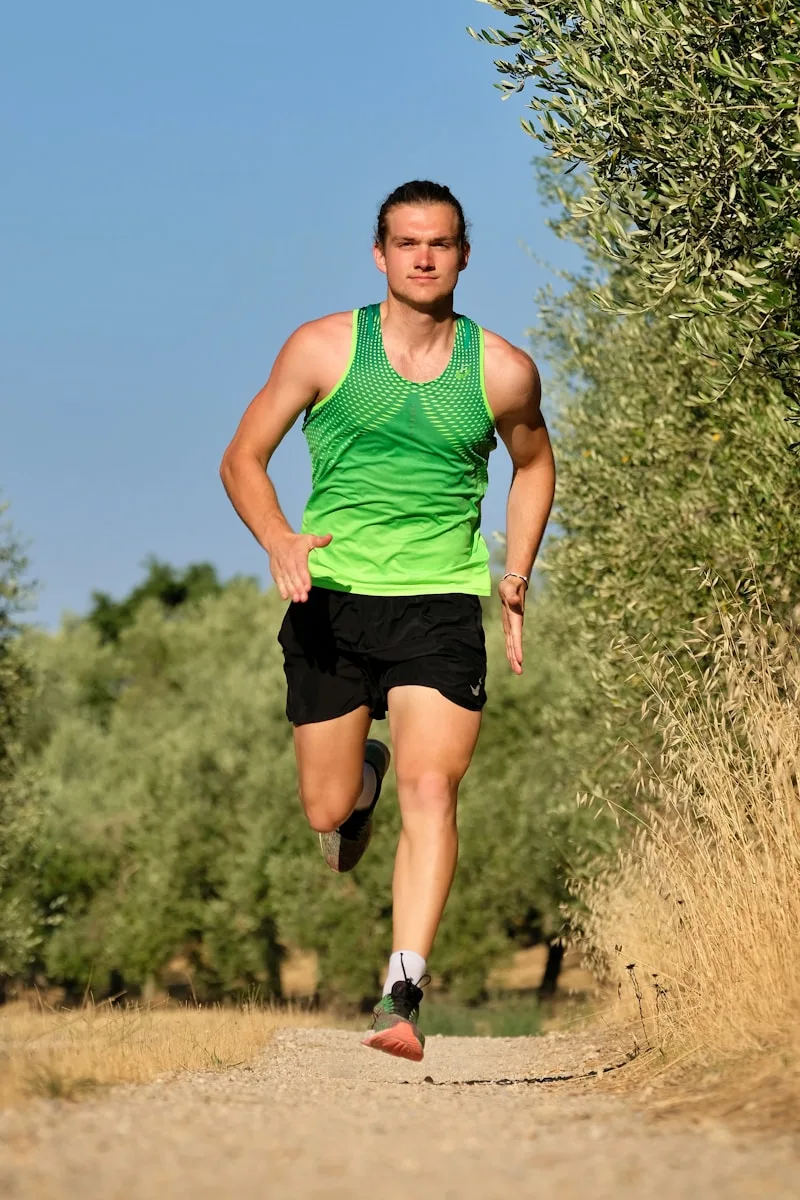
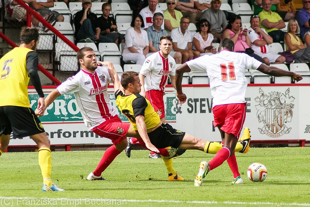
Runner clutching lower leg in pain after a calf strain“I wasn’t even sprinting…” It’s one of the most common stories I hear in the clinic. “I pushed off to chase the football” “I accelerated after my dog.” “I was only halfway through my run.” “It felt like someone kicked me in the calf.” The diagnosis is usually the same: a calf strain.

In sporting circles, particularly AFL, this injury has become so common that it has earned its own nickname: “Old Man Calf.” The term is tongue in cheek, but the injury certainly isn’t. Calf strains are one of the most frustrating soft-tissue injuries affecting middle-aged athletes and recreational runners. They also have a well documented habit of recurring when they aren’t managed properly.

As someone who treats these injuries regularly, and who has experienced a few myself, I know how debilitating and frustrating they can be. The encouraging part is that they’re also one of the more preventable injuries once you understand why they happen.

Why does it happen?

Age itself isn’t really the problem. What ageing does is create a perfect storm of small physiological changes that gradually chip away at the calf’s ability to tolerate high speed loading.

These changes include a reduction in muscle mass, known as sarcopenia, driven largely by the progressive loss of motor neurons and the muscle fibres they supply (Larsson et al., 2019; Nishikawa et al., 2021). Achilles tendon stiffness also appears to decline with age. Research using ultrasound and biofeedback found that older adults produced up to 44% less tendon stiffness than younger adults at matched levels of muscle activation. That alters how efficiently force transfers from muscle to bone during push-off (Gray et al., 2024). Interestingly, when load rather than effort is matched, some studies find no real difference in tendon stiffness between younger and older adults. That suggests a good portion of this “stiffness loss” is really a downstream effect of age-related strength loss rather than the tendon tissue itself deteriorating (Jakubowski et al., 2024). Either way, the practical implication is the same: rebuilding strength is central to rebuilding tendon function. Alongside these structural changes, recovery between training sessions slows, maximal strength and power decline, and most people simply stop exposing their calves to genuine sprinting on a regular basis.

None of these changes is dramatic on its own. Together, though, they mean the calf has less “reserve capacity” available when a sudden, explosive movement is demanded of it. That’s why so many people tear a calf doing something they’ve done hundreds of times before without issue.

Why AFL players seem particularly vulnerable

If you’ve watched AFL over the past decade, you’ve probably heard the commentary: “another calf injury,” with veteran players seeming especially susceptible. This isn’t coincidence.

Older AFL players typically retain excellent aerobic fitness and game awareness. But they’re still required to produce repeated maximal accelerations, decelerations and changes of direction, and these explosive actions place enormous force through the calf complex. Data from the AFL’s Soft Tissue Injury Registry back this up. An early analysis of muscle strains across the competition found that age was an independent risk factor for calf strains even after adjusting for injury history, while it wasn’t a risk factor for quadriceps strains. That suggests there’s something specific about how the calf tolerates ageing under repeated high-speed loading (Orchard, 2001). A later 23-year cohort of more than 3,600 muscle strains across the AFL confirmed the pattern, with each additional year of age increasing the odds of a calf strain by around 60% (Orchard et al., 2020). Older players who do sustain a calf strain also tend to take longer to recover (Gray et al., 2025). A history of calf strain combined with older age also substantially raises the risk of a further injury within the following two seasons (Green et al., 2020).

Elite sport doesn’t create this vulnerability; it simply exposes it more publicly.

Recreational runners aren’t immune

You don’t need to play AFL to be at risk. Many of the calf tears seen in clinic occur in park runners, masters athletes, weekend footballers, tennis and pickleball players, golfers walking steep courses, and parents chasing kids around the backyard. Pickleball in particular has seen a sharp rise in soft tissue injuries as participation among older adults has grown, with calf strains featuring among the most commonly recorded injuries at one US orthopaedic centre (Herzberg et al., 2025).

The common thread in all these cases is an athlete who remains active, but who isn’t regularly exposing their calves to high-speed loading. Your cardiovascular fitness might still be excellent. Your calf’s capacity to absorb a sudden sprint or lunge may not be.

Which muscle actually tears?
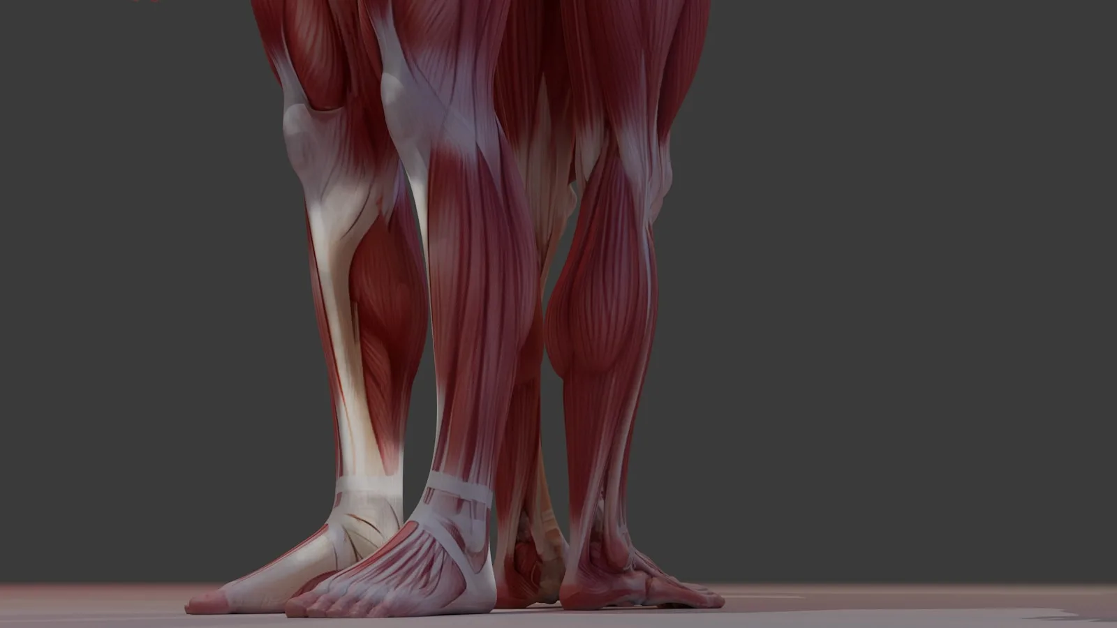
Detailed anatomical view of the calf musclesMost people simply say they’ve “done their calf,” but several distinct structures can be involved, and it’s worth knowing the difference.

Gastrocnemius is the larger, more superficial of the two major calf muscles. It’s typically injured during explosive acceleration, jumping or sprinting, and pain is often felt high in the calf and occurs suddenly. In a 10-year cohort of elite AFL players, gastrocnemius injuries most commonly affected the medial head (78% of cases), with acceleration being the single most common mechanism of injury (Gray et al., 2025).

Soleus sits deeper and functions more as an endurance muscle. It’s commonly injured during running or prolonged loading, and pain is usually felt lower in the calf, often starting as tightness before becoming more obviously painful. In the same injury registry, soleus injuries were actually more prevalent overall than gastrocnemius injuries, accounting for around 85% of all calf strains in elite AFL players. They were also far more likely to recur (Green et al., 2019). Because soleus symptoms tend to develop more gradually, they’re often under-recognised in older runners until the injury is well established.

Why do calf tears keep coming back?

This is probably the biggest source of frustration for patients. Many people feel “pretty good” after three to four weeks: they can walk, they can cycle, and they might even jog comfortably. Unfortunately, healing tissue isn’t the same as fully restored tissue.

The evidence on recurrence is sobering. In elite AFL players, a prior calf strain increased the risk of a further injury several-fold. Older age independently increased that risk further still (Green et al., 2020). A separate analysis of over 3,600 muscle strains across 23 AFL seasons found that the risk of recurrence stays elevated for around 15 weeks after return to play. It also found that a recent history of the same injury was, by a wide margin, the strongest predictor of injuring it again (Orchard et al., 2020). Put simply, unless strength, endurance, plyometric capacity and sprint exposure are specifically rebuilt, the calf returns to sport with reduced capacity, and eventually something gives again.

Can you prevent “Old Man Calf”?

The encouraging answer is yes. While age can’t be changed, capacity can.

Heavy calf strength. Strong calves tolerate greater force before they fail. Heavy seated and standing calf raises should be part of long-term training, not something reserved for rehab after an injury has already occurred. Eccentric loading in particular has been shown to drive meaningful adaptations in tendon properties. It may be especially well suited to older adults, since it can achieve those adaptations at comparatively lower relative demands than other contraction types (Quinlan et al., 2019).

Plyometric training. Skipping, hopping and bounding, performed in small, consistent doses, help the calf improve its ability to store and release elastic energy efficiently.
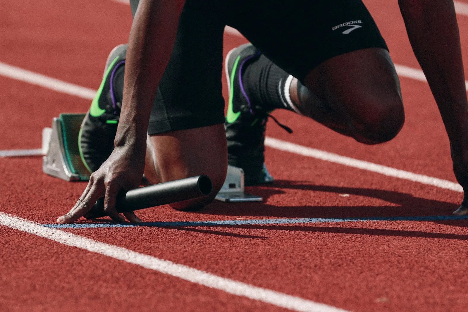
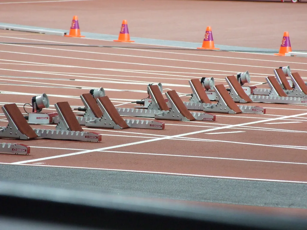
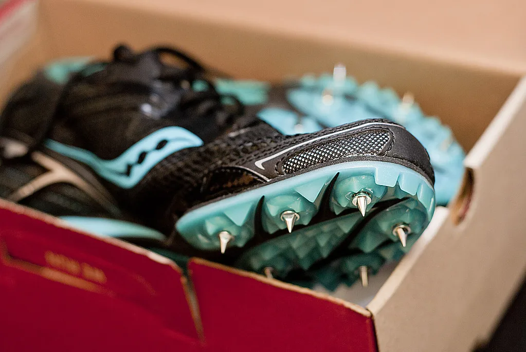
Runner in starting blocks preparing to sprintSprint exposure. This is perhaps the most overlooked factor of all. Many recreational runners train at a single, moderate pace and then, once every few months, need to sprint for a bus, a ball or a toddler heading for the road. That’s the equivalent of expecting a shoulder to throw a ball at full pace without ever having practised it. Regular controlled strides, accelerations and genuinely faster running help maintain the calf’s resilience to sudden demand.

Consistent training volume. Sharp spikes in training load remain one of the most consistently identified injury risk factors. In professional footballers, a week of unusually high external workload, particularly involving high-intensity accelerations and decelerations, regularly preceded calf strain injuries. Injured players were, on average, older than their uninjured teammates too (Soler et al., 2024). The calf tends to prefer gradual, planned increases over heroic weekends bolted onto weeks of inactivity.

Recovery. Sleep, nutrition and ongoing strength training all matter more after 40, simply because recovery isn’t as forgiving as it once was.

Don’t ignore persistent calf tightness

One of the most common mistakes I see is assuming every tight calf just needs stretching. Persistent tightness can actually represent residual weakness, reduced tendon capacity, neural fatigue, or an incompletely healed muscle injury. Stretching alone rarely resolves any of these underlying issues.

A proper assessment should identify why the calf feels tight in the first place. Often, the answer is that it needs to get stronger, not longer.

When should you seek help?

It’s worth having the injury assessed if you experience sudden pain with a popping sensation, difficulty pushing up onto your toes, swelling or bruising, repeated calf strains, or calf tightness that never quite seems to resolve.

A detailed examination can usually determine which structure has been injured, whether imaging is required, and, most importantly, what needs to be rebuilt before you return to running or sport.

The bottom line

“Old Man Calf” might sound like a joke, but it reflects a genuine physiological phenomenon. As we age, our calves gradually lose reserve capacity. And without ongoing strength, power and speed exposure, they’re regularly asked to perform tasks they simply haven’t been prepared for.

Fortunately, this isn’t inevitable. With the right combination of strength training, progressive running, plyometrics and intelligent rehabilitation, many athletes continue running, competing and enjoying sport well into their 50s, 60s and beyond. Age may increase the risk, but it doesn’t have to define your future. If you have struggled (like I have) with calf tears derailing your activity participation, or you fear the return to running stage, book in with one of our physios today to get you back to your best! 

Until next time, Praxis What You Preach 

📍 Clinics in Teneriffe, Buranda, and Carseldine

💪 Trusted by athletes. Backed by evidence. Here for everyone.

Related Articles
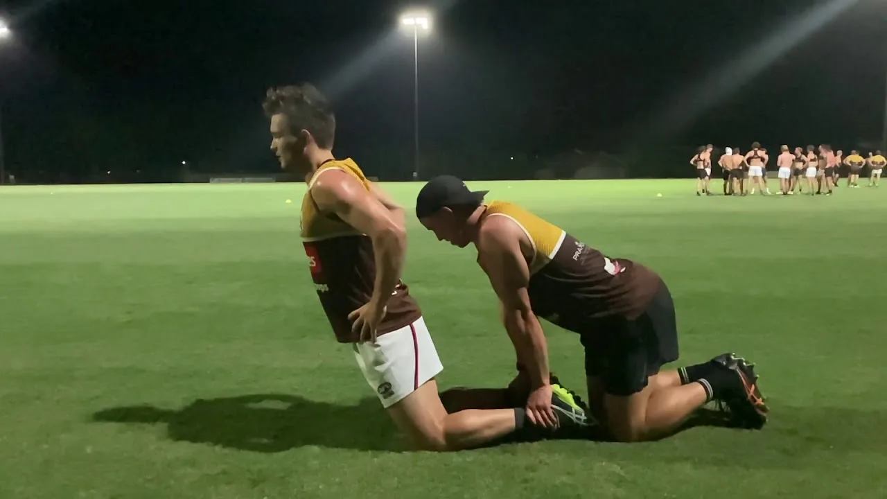
Nordic Curls and the Copenhagen Plank: Two Exercises That Could Save Your Season
Two simple exercises, backed by strong evidence, for cutting your hamstring and groin injury risk down significantly.

Shin Splints: Causes, Treatment & How to Get Back to Running Stronger
That nagging ache down your shin isn’t something to just run through. Here’s what’s actually going on, and how to fix it.

Plantar Fasciopathy: Understanding how to heal your heel pain

Heel pain first thing in the morning is one of the most common, and most misunderstood, overuse injuries we see.

References

  1. Larsson L, Degens H, Li M, et al. Sarcopenia: Aging-Related Loss of Muscle Mass and Function. Physiol Rev. 2019;99(1):427-511. DOI
  2. Nishikawa H, Fukunishi S, Asai A, et al. Pathophysiology and mechanisms of primary sarcopenia (Review). Int J Mol Med. 2021;48(2). DOI
  3. Gray AJ, Krupenevich RL, Batsis JA, Sawicki GS, Franz JR. Reduced Achilles tendon stiffness in aging associates with higher metabolic cost of walking. J Appl Physiol. 2024;137(6):1541-1548. DOI
  4. Jakubowski KL, Ludvig D, Lee SSM, Perreault EJ. Aging Does Not Alter Ankle, Muscle, and Tendon Stiffness at Low Loads Relevant to Stance. Ann Biomed Eng. 2024;52(9):2556-2568. DOI
  5. Orchard JW. Intrinsic and extrinsic risk factors for muscle strains in Australian football. Am J Sports Med. 2001;29(3):300-303. DOI
  6. Orchard JW, Chaker Jomaa M, Orchard JJ, et al. Fifteen-week window for recurrent muscle strains in football. Br J Sports Med. 2020;54(18):1103-1107. DOI
  7. Gray C, Pizzari T, Murphy MC, et al. Gastrocnemius Muscle Strain Injury Characteristics in Elite Male Australian Football Players: A 10-Year Longitudinal Cohort Study. J Orthop Sports Phys Ther. 2025;55(10):681-688. DOI
  8. Green B, Lin M, McClelland JA, Semciw AI, Schache AG, Rotstein AH, Cook J, Pizzari T. Return to Play and Recurrence After Calf Muscle Strain Injuries in Elite Australian Football Players. Am J Sports Med. 2020;48(13):3306-3315. DOI
  9. Herzberg SD, Bowman EN, Hill KL. Evaluation of Pickleball-Related Injuries at a Single Institution From 2017 to 2022. Orthop J Sports Med. 2025;13(2). DOI
  10. Green B, Lin M, Schache AG, McClelland JA, Semciw AI, Rotstein A, Cook J, Pizzari T. Calf muscle strain injuries in elite Australian Football players: A descriptive epidemiological evaluation. Scand J Med Sci Sports. 2020;30(1):174-184. DOI
  11. Quinlan JI, Narici MV, Reeves ND, Franchi MV. Tendon Adaptations to Eccentric Exercise and the Implications for Older Adults. J Funct Morphol Kinesiol. 2019;4(3):60. DOI
  12. Soler A, Agulló F, Hernández-Dávó J, Raya-González J, Del Coso J, González-Ródenas J, Moreno-Pérez V. Influence of the External Workload on Calf Muscle Strain Injuries in Professional Football Players: A Pilot Study. Sports Health. 2024;17(1):175-182. DOI
The Single-Leg Squat Test: The Hip Control Test That Predicts Your Next Knee Injury

The Single-Leg Squat Test: The Hip Control Test That Predicts Your Next Knee Injury

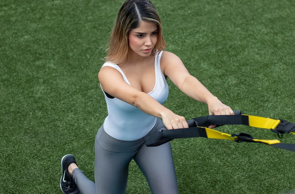
Athlete performing a lunge, loading the hip and gluteal muscles

Hip and gluteal control rarely gets tested properly before return to sport, largely because it’s easy to mistake for something else. An athlete with poor hip control can usually still run in a straight line without pain. That’s exactly why the deficit tends to only show up later, in a knee, a groin, or the same hip again, once the demands of cutting and landing expose what straight-line running never did. The single-leg squat and step-down tests are two of the simplest, best-evidenced ways to catch that gap early.

WHAT THE TEST ACTUALLY MEASURES

Both tests are performed standing on one leg, either squatting down as far as controlled technique allows, or stepping down from a small box or step. What’s being assessed isn’t just how deep the movement goes, but the quality of control through it. Does the knee track over the foot or collapse inward? Does the pelvis stay level or drop on the unsupported side? Does the whole movement look stable or wobbly? These patterns are driven largely by hip abductor and external rotator strength, particularly gluteus medius, which is why the test is as much a hip test as it is a knee test.

A 2011 study by Poulsen and James, published in Physiotherapy Theory and Practice, examined the reliability and validity of clinical evaluation of the single-leg squat and found it to be a reliable, valid way for clinicians to assess lower limb control, even when scored visually rather than with laboratory equipment. That’s a meaningful finding, because it means this test doesn’t require expensive gear to be clinically useful, just a trained eye and a consistent scoring approach.

WHY TESTING BEATS GUESSING

Hip control deficits are easy to miss because they often don’t cause pain in the hip itself. An athlete can walk, jog, and even sprint in a straight line with poor gluteal control and feel completely normal, right up until a cutting or landing task demands single-leg stability under load. Without a specific test for this, it simply doesn’t get picked up before return to sport.

This mirrors the broader return-to-sport literature: a 2016 Delaware-Oslo cohort study by Grindem and colleagues found an 84% lower reinjury rate in athletes cleared using objective discharge criteria rather than time alone, and a related study by Kyritsis and colleagues found a fourfold higher reinjury risk in athletes who didn’t meet full discharge criteria. The single-leg squat and step-down tests apply that same principle specifically to the hip and pelvis, an area that’s otherwise very easy to clear on the basis of “no pain” alone.

THE KINETIC CHAIN COST OF CUTTING CORNERS

Poor hip control is one of the clearest examples of a kinetic chain problem in sports medicine, because the knee, not the hip, is usually where the consequences show up. When the pelvis drops and the knee collapses inward during single-leg loading, it changes the angle and force through the knee joint on every step, cut, and landing. That’s a pattern strongly implicated in patellofemoral pain and non-contact ACL injury. The same redistribution effect is well documented after other underprepared joints return to sport. A 2016 systematic review by Wiggins and colleagues found that athletes returning to sport after ACL reconstruction reinjured the same knee and the opposite knee at almost identical rates. That’s a pattern consistent with hip control deficits driving risk into the knee on both sides rather than staying contained at the hip.

Left unaddressed over years of training and competition, this pattern of poor load control through the hip and knee is also implicated in the gradual development of joint degeneration. The joint surfaces end up repeatedly loaded in ways they weren’t designed for. Confirming genuine hip control before returning to full training is one of the more practical ways to interrupt that pattern. It stops the deficit becoming a long-term problem rather than a single injury.

WHAT WE DO AT PRAXIS

We use single-leg squat and step-down assessment alongside gluteal strength testing and landing mechanics screening. Together, these confirm the hip is genuinely controlling load before we clear an athlete back to cutting, jumping, or contact sport. If you’re recovering from a hip, groin, or knee injury, or you simply want to know how your hip control measures up, book a return-to-sport assessment with our team.

Until next time, Praxis What You Preach…

📍 Clinics in Teneriffe, Woolloongabba, and Carseldine

💪 Trusted by athletes. Backed by evidence. Here for everyone.

Related Articles

The Single Leg Squat

A closer look at the single leg squat, one of the most useful clinical tests of hip and knee control.

Unilateral vs Bilateral Training: Part 1 - Your Comparative Guide

How single-leg training builds the hip and knee control that bilateral training alone can miss.

Return to Sport Testing: The Complete Guide

One evidence-based capacity test for every major joint, and why testing beats guessing.

References

Poulsen DR, James CR. Concurrent validity and reliability of clinical evaluation of the single leg squat. Physiotherapy Theory and Practice. 2011;27(8):586-594.

Grindem H, Snyder-Mackler L, Moksnes H, Engebretsen L, Risberg MA. Simple decision rules can reduce reinjury risk by 84% after anterior cruciate ligament reconstruction: the Delaware-Oslo ACL cohort study. British Journal of Sports Medicine. 2016;50(13):804-808.

Kyritsis P, Bahr R, Landreau P, Miladi R, Witvrouw E. Likelihood of ACL graft rupture: not meeting six clinical discharge criteria before return to sport is associated with a four times greater risk of rupture. British Journal of Sports Medicine. 2016;50(15):946-951.

Wiggins AJ, Grandhi RK, Schneider DK, Stanfield D, Webster KE, Myer GD. Risk of secondary injury in younger athletes after anterior cruciate ligament reconstruction: a systematic review and meta-analysis. American Journal of Sports Medicine. 2016;44(7):1861-1876.

The Single-Leg Hop Test: Why Most Ankle Sprains Are Cleared Before They’re Actually Ready

The Single-Leg Hop Test: Why Most Ankle Sprains Are Cleared Before They’re Actually Ready

Trail runner landing on one leg, the movement pattern assessed by ankle hop testing

Ankle sprains are treated as minor injuries more often than almost anything else in sport, which is part of the problem. Most people are walking normally within days and back at light training within a couple of weeks. That’s often long before the ankle has actually regained the strength and control needed to land, cut, or change direction without giving way again. A single-leg hop test is one of the more practical ways to check that control has genuinely returned before an athlete finds out the hard way.

WHAT THE TEST ACTUALLY MEASURES

The test itself is straightforward. Standing on one leg, the athlete hops forward as far as possible and lands under control on the same foot, without the free leg touching down or the landing foot needing to step to regain balance. Distance is measured and compared side to side. The quality of the landing, whether it’s stable and controlled or wobbly and compensated, is assessed alongside the raw number.

A 2009 study by Caffrey, Docherty, Schrader, and Klossner, published in the Journal of Orthopaedic and Sports Physical Therapy, examined four different single-limb hopping tests in people with functional ankle instability and found that hopping tests were able to detect functional performance deficits that simpler balance and range-of-motion measures missed. The general benchmark used across the hop testing literature, consistent with lower limb testing more broadly, is a Limb Symmetry Index of at least 90% between the injured and uninjured ankle before returning to unrestricted sport.

WHY TESTING BEATS GUESSING

Because ankle sprains are so common and usually resolve pain quickly, they’re one of the injuries most likely to be self-managed without any formal testing before return to sport. That’s exactly why they’re also one of the most likely injuries to recur, with a well-established pattern where an initial sprain, especially if returned to sport too early, tends to be followed by a second and third, eventually settling into what’s recognised clinically as chronic ankle instability.

The broader evidence for objective testing over time-based clearance is consistent across joints. A 2016 Delaware-Oslo cohort study by Grindem and colleagues found an 84% lower reinjury rate in athletes cleared using objective discharge criteria rather than time alone, and a related study by Kyritsis and colleagues found a fourfold higher reinjury risk in athletes who didn’t meet full discharge criteria before returning to sport. A hop test applies exactly that principle to the ankle: confirm the capacity is actually there, rather than assuming it from the absence of pain.

THE KINETIC CHAIN COST OF CUTTING CORNERS

An ankle that returns to sport with reduced control rarely stays an isolated ankle problem. Athletes with chronic ankle instability commonly develop compensatory movement patterns up the chain. They alter knee and hip mechanics during landing and cutting to protect the unstable ankle, which shifts injury risk upward rather than removing it. This same load-redistribution pattern is well documented after other undertested joints return to sport: a 2016 systematic review by Wiggins and colleagues found that athletes returning to sport after ACL reconstruction reinjured the same knee and the opposite knee at almost identical rates. This illustrates how an underprepared joint changes the loading pattern of everything connected to it, not just itself.

Each recurrent ankle sprain also tends to leave the joint a little less stable than before. That compounds the risk of the next one and increases the long-term likelihood of chronic instability and early degenerative change in the joint. Confirming hop test performance before return to sport is one of the more effective ways to break that cycle rather than let it repeat every season.

WHAT WE DO AT PRAXIS

We use single-leg hop testing alongside balance and proprioception assessment and sport-specific cutting drills. Together, these confirm an ankle is genuinely ready for full training, not just pain-free at walking pace. If you’re recovering from an ankle sprain, or you’ve had more than one, book a return-to-sport assessment with our team.

Until next time, Praxis What You Preach…

📍 Clinics in Teneriffe, Woolloongabba, and Carseldine

💪 Trusted by athletes. Backed by evidence. Here for everyone.

Related Articles

Ankle Sprains: Don't Let a Simple Injury Turn into a Long-Term Problem

Why ankle sprains are so often undertreated, and what evidence-based rehab actually looks like.

Achilles Tendinopathy: How to treat your Achilles Pain

How reduced ankle and calf capacity can contribute to Achilles pain, and the evidence-based approach to treating it.

Return to Sport Testing: The Complete Guide

One evidence-based capacity test for every major joint, and why testing beats guessing.

References

Caffrey E, Docherty CL, Schrader J, Klossner J. The ability of 4 single-limb hopping tests to detect functional performance deficits in individuals with functional ankle instability. Journal of Orthopaedic and Sports Physical Therapy. 2009;39(11):799-806.

Grindem H, Snyder-Mackler L, Moksnes H, Engebretsen L, Risberg MA. Simple decision rules can reduce reinjury risk by 84% after anterior cruciate ligament reconstruction: the Delaware-Oslo ACL cohort study. British Journal of Sports Medicine. 2016;50(13):804-808.

Kyritsis P, Bahr R, Landreau P, Miladi R, Witvrouw E. Likelihood of ACL graft rupture: not meeting six clinical discharge criteria before return to sport is associated with a four times greater risk of rupture. British Journal of Sports Medicine. 2016;50(15):946-951.

Wiggins AJ, Grandhi RK, Schneider DK, Stanfield D, Webster KE, Myer GD. Risk of secondary injury in younger athletes after anterior cruciate ligament reconstruction: a systematic review and meta-analysis. American Journal of Sports Medicine. 2016;44(7):1861-1876.

The Biering-Sørensen Test: The Trunk Endurance Number Your Back Rehab Is Probably Skipping

The Biering-Sørensen Test: The Trunk Endurance Number Your Back Rehab Is Probably Skipping

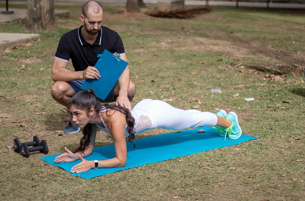
Physiotherapist coaching a plank exercise for trunk extensor endurance

Low back pain is unusual among sporting injuries in that it rarely announces a clear moment of “reinjury” the way a hamstring or ankle does. Instead it tends to creep back in: a bit stiffer this week, a bit more guarded the next. Before long, an athlete is training below their capacity without anyone quite deciding they should be. The Biering-Sørensen test is one of the few simple, well-validated tools for putting a number on trunk extensor endurance before that pattern sets in.

WHAT THE TEST ACTUALLY MEASURES

The test is performed lying face down with the pelvis and legs fixed on a table, the upper body unsupported and held horizontal, in line with the legs. The time the position can be held before form breaks down is recorded. It’s a direct measure of isometric endurance in the back extensors. These are the muscles that keep the trunk upright and controlled during running, lifting, and repeated bending and twisting under fatigue.

The test was first described by Biering-Sørensen in a 1984 study published in Spine, which followed working adults for a year and found that a shorter holding time was a significant predictor of who would go on to develop low back trouble. A 1999 study by Latimer and colleagues, published in the same journal, confirmed the test’s reliability and its ability to discriminate between people with and without low back pain, reporting good to excellent reliability across asymptomatic and symptomatic groups.

WHY TESTING BEATS GUESSING

Because low back pain so often fluctuates rather than resolving in a straight line, athletes and clinicians can end up treating “no pain today” as the signal to return to full training, without ever confirming that trunk endurance has actually been restored. That’s a meaningful gap: an athlete can be pain-free at rest and still fatigue through their back extensors well before the end of a training session or a match, at which point technique breaks down and injury risk rises.

The same principle holds across other joints in the return-to-sport literature. A 2016 Delaware-Oslo cohort study by Grindem and colleagues found an 84% lower reinjury rate in athletes cleared using objective discharge criteria rather than time alone. A related study by Kyritsis and colleagues found a fourfold higher reinjury risk in athletes who didn’t meet full discharge criteria. Trunk endurance testing applies the same logic to the lower back: a number that has to be met, not a symptom that simply has to be absent.

THE KINETIC CHAIN COST OF CUTTING CORNERS

A fatiguing back extensor doesn’t fail in isolation. As trunk endurance drops, athletes typically lose postural control earlier in a session. That changes how load is transferred down into the hips and up through the shoulders during running, lifting, and repeated bending. This kind of load redistribution, where an underprepared segment pushes risk onto the joints around it, is well documented elsewhere in return-to-sport research. A 2016 systematic review by Wiggins and colleagues found that athletes returning to sport after ACL reconstruction reinjured the same knee and the opposite knee at almost identical rates. This illustrates how an untested weak link doesn’t just risk itself: it changes the loading pattern of everything connected to it.

Biering-Sørensen’s own findings make the long-term stakes clear: reduced back extensor endurance was directly predictive of who developed low back trouble over the following year. Confirming trunk endurance before returning to full training or competition is one of the more practical ways to interrupt that pattern. It stops a recurring, low-grade problem from following an athlete from one season into the next.

WHAT WE DO AT PRAXIS

We use the Biering-Sørensen test alongside movement screening and graded loading through the trunk and hips. Together, these confirm the back is genuinely ready for full training, not just quiet at rest. If you’re managing low back pain or returning from a back injury, book a return-to-sport assessment with our team.

Until next time, Praxis What You Preach…

📍 Clinics in Teneriffe, Woolloongabba, and Carseldine

💪 Trusted by athletes. Backed by evidence. Here for everyone.

Related Articles

Lower Back Pain

What the evidence says about sciatica, and why the outlook is usually far better than it feels.

Pilates & Back Pain Rehab (Part 3 of 3): How Much Is Enough?

How much rehab is actually enough when it comes to back pain, and how to know when you’ve done it.

Return to Sport Testing: The Complete Guide

One evidence-based capacity test for every major joint, and why testing beats guessing.

References

Biering-Sørensen F. Physical measurements as risk indicators for low-back trouble over a one-year period. Spine. 1984;9(2):106-119.

Latimer J, Maher CG, Refshauge K, Colaco I. The reliability and validity of the Biering-Sørensen test in asymptomatic subjects and subjects reporting current or previous nonspecific low back pain. Spine. 1999;24(20):2085-2090.

Grindem H, Snyder-Mackler L, Moksnes H, Engebretsen L, Risberg MA. Simple decision rules can reduce reinjury risk by 84% after anterior cruciate ligament reconstruction: the Delaware-Oslo ACL cohort study. British Journal of Sports Medicine. 2016;50(13):804-808.

Kyritsis P, Bahr R, Landreau P, Miladi R, Witvrouw E. Likelihood of ACL graft rupture: not meeting six clinical discharge criteria before return to sport is associated with a four times greater risk of rupture. British Journal of Sports Medicine. 2016;50(15):946-951.

Wiggins AJ, Grandhi RK, Schneider DK, Stanfield D, Webster KE, Myer GD. Risk of secondary injury in younger athletes after anterior cruciate ligament reconstruction: a systematic review and meta-analysis. American Journal of Sports Medicine. 2016;44(7):1861-1876.

The Adductor Squeeze Test: The Groin Strength Number That Predicts Reinjury

The Adductor Squeeze Test: The Groin Strength Number That Predicts Reinjury

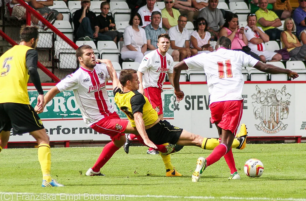
Football player lunging for the ball, loading the hip adductors

Groin and adductor strains are among the most persistent injuries in football codes. One of the biggest reasons is that they’re often cleared to return based on pain settling during light activity, well before the adductor can produce the forceful, high-speed contraction needed to kick, cut, or change direction at pace. The adductor squeeze test gives us a way to measure that gap directly, rather than guess at it.

WHAT THE TEST ACTUALLY MEASURES

The test is performed lying on your back with the hips and knees bent, squeezing a pressure cuff, dynamometer, or the assessor’s hands between the knees as hard as possible for a few seconds. It’s a simple, quick measure of maximal isometric hip adductor strength. It can be performed in both a short-lever position (knees bent) and a long-lever position (legs straighter), which stress the adductors slightly differently.

A 2019 prospective study by Moreno-Pérez and colleagues, published in Physical Therapy in Sport, followed 71 elite football players across a season and found that low adductor squeeze strength was directly associated with a higher probability of sustaining a groin injury. Players with maximal isometric adductor force below a specific threshold had a substantially higher chance of injury, and the association held even after accounting for body mass. That’s a genuinely useful, actionable number, not just a general impression of “tightness” or discomfort.

WHY TESTING BEATS GUESSING

Groin pain is notoriously slow and non-linear to resolve, and athletes and clinicians alike can be tempted to return to sport once pain-free walking, jogging, and even straight-line running have been achieved. The trouble is that a forceful cut, a kick, or a change of direction loads the adductors in a completely different way, at much higher speed and force, and that’s exactly where an undertested adductor tends to fail.

This mirrors return-to-sport research from other joints: a 2016 Delaware-Oslo cohort study by Grindem and colleagues found an 84% lower reinjury rate in athletes cleared using objective discharge criteria rather than time alone. A related study by Kyritsis and colleagues found a fourfold higher reinjury risk in athletes who didn’t meet full discharge criteria before returning. The adductor squeeze test is the groin’s equivalent of those objective markers: a number that has to be met, not a feeling that has to pass.

THE KINETIC CHAIN COST OF CUTTING CORNERS

Weak adductors don’t just risk a repeat groin strain. The adductors work closely with the hamstrings and the deep hip stabilisers to control the leg during sprinting and cutting. A deficit in one muscle group often shows up as compensatory overload in a neighbouring one. That’s part of why hamstring and groin injuries so often cluster together in the same athletes and the same seasons. The broader principle, that an undertested muscle group pushes load onto whatever is next in the chain, is well documented in ACL research. A 2016 systematic review by Wiggins and colleagues found that athletes returning to sport after ACL reconstruction re-injured the same knee and the opposite knee at almost identical rates. This illustrates how an underprepared joint or muscle group redistributes risk rather than eliminating it.

Chronic or recurrent groin pain, sometimes progressing to what’s described as athletic pubalgia, also becomes harder to fully resolve the more times it recurs. Each recurrence leaves more secondary compensation patterns to untangle. Confirming adequate adductor strength before full return to sport is one of the more effective ways to keep a single strain from becoming a recurring, season-defining problem.

WHAT WE DO AT PRAXIS

We use the adductor squeeze test alongside Copenhagen plank progressions and sport-specific cutting and kicking drills. Together, these confirm the groin is genuinely ready for full training, not just comfortable at jogging pace. If you’re managing groin or adductor pain, book a return-to-sport assessment with our team.

Until next time, Praxis What You Preach…

📍 Clinics in Teneriffe, Woolloongabba, and Carseldine

💪 Trusted by athletes. Backed by evidence. Here for everyone.

Related Articles

Chronic Groin Pain (Athletic Pubalgia)

Understanding chronic groin pain and athletic pubalgia, and the evidence-based approach to resolving it.

Nordic Curls and the Copenhagen Plank: Two Exercises That Could Save Your Season

The two exercises with the strongest evidence behind them for reducing hamstring and groin strain risk.

Return to Sport Testing: The Complete Guide

One evidence-based capacity test for every major joint, and why testing beats guessing.

References

Moreno-Pérez V, Travassos B, Calado A, Gonzalo-Skok O, Del Coso J, Mendez-Villanueva A. Adductor squeeze test and groin injuries in elite football players: a prospective study. Physical Therapy in Sport. 2019;37:54-59.

Grindem H, Snyder-Mackler L, Moksnes H, Engebretsen L, Risberg MA. Simple decision rules can reduce reinjury risk by 84% after anterior cruciate ligament reconstruction: the Delaware-Oslo ACL cohort study. British Journal of Sports Medicine. 2016;50(13):804-808.

Kyritsis P, Bahr R, Landreau P, Miladi R, Witvrouw E. Likelihood of ACL graft rupture: not meeting six clinical discharge criteria before return to sport is associated with a four times greater risk of rupture. British Journal of Sports Medicine. 2016;50(15):946-951.

Wiggins AJ, Grandhi RK, Schneider DK, Stanfield D, Webster KE, Myer GD. Risk of secondary injury in younger athletes after anterior cruciate ligament reconstruction: a systematic review and meta-analysis. American Journal of Sports Medicine. 2016;44(7):1861-1876.