Stingers AKA Neural traction injuries

Stingers AKA Neural traction injuries

SUMMARY:

  • Stingers are essentially a tractioning of the neural system
  • This can cause pain, movement and sensation changes
  • Chronic traction to the nervous system can have a cumulative effect on nerve function
  • The more damage to the nerve, the more serious the outcome
  • We shouldn’t be as dismissive of “stingers”, particularly if they are recurrent
  • Physiotherapy has a role to play

STINGERS:

Stingers have been the catch cry of many contact sports over the years. Often dismissed as ‘just a stinger’, trauma to the nervous system should probably be taken a little more seriously, as we delve into detail today.

Stingers are most commonly experienced in contact sports whereby the shoulder of a players is forcefully depressed, as experienced with a tackle in NRL or union. A large range of motion over a short period of time can result in a ‘traction’ of the Brachial Plexus (a network of nerves formed from exiting branches of the spinal cord in the neck that transverse to the shoulder and arm). Thus network of nerves sends signals from your spinal cord to your shoulder, arm and hand and thereby providing feeling and movement to these regions.

BACKGROUND:

To understand a stinger injury, a clear understanding of nerves and nerve related injuries is required. A nerve is an enclosed, cable-like bundle of nerve fibres called axons, in the peripheral nervous system. A nerve provides a common pathway for the electrochemical nerve impulses that provide a number of functions, including getting our muscles to move!

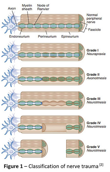
Seddon and Sunderland present a five-grade classification scale for nerve related injuries[1-4]. Figure 1 illustrates the physiological changes that occur through each grade of injury. Essentially it outlines the greater amount of disruption to the anatomy of the nerve.

The more damage to the nerve, the more serious the outcome. Table 1 outlines the three different grades of stinger injuries.[4, 5] The most common stinger is a grade 1 injury, which represents a neurapraxia, or nerve stretch injury, without axonal disruption.[4] In an acute setting, this can result in motor and sensory loss/changes, which usually resolve within minutes.[1, 4] Grade 2 and 3 involve a higher degree of nerve injury, usually involving a crush, transection or compression mechanism.[1]

Chronic traction to the nervous system can have a cumulative effect on nerve function. This is termed “chronic stinger syndrome” and represents a distinct entity from acute stingers that may reflect long- standing structural changes of the subaxial spinal canal and chronic irritation/degeneration of the exiting nerve root complex.[4, 6]. This sounds complex but essentially means

Mid Potion Achilles Tendinopathy Location

A Clinical Example from Zac

“During a Gridiron match, a player was injured whilst making a tackle. I reviewed the player on field and he was unable to utilise his right upper limb (full paralysis) from shoulder down to his hand. The player was removed from the field immediately to be further assessed and monitored. A complete neurovascular assessment was performed, assessing motor function/strength, sensation, reflexes, and vascular status, as well was the cervical spine. Motor and sensation changes were the only deficits noted and were reviewed frequently. After roughly five minutes, the player demonstrated full upper limb motor strength and sensation, with nil lingering symptoms. In collaboration with the patient, it was decided he would return to match play immediately. The patient was monitored throughout the game and reported no further symptoms.”

Figure 2 shows a proposed decision tree when managing stinger injuries.[4] This clinical example outline above fits the Grade 1 Mild category as he was able to return to competition with nil lingering symptoms. Despite the lack of symptoms during the game, it is recommended the patient be reviewed again both after the game and weekly for two weeks to ensure a full resolution of symptoms.[4, 7]

The role for neural mobilisation?

Current non-surgical management involves rest, pain control and resistance training[4]. Though not explored within the literature, neural mobilization may have an important role in patients with persistent symptoms, such as Grade 1 moderate to severe, and more recurrent neuropraxias. Though not assessed in this specific population, there is evidence for neural tissue management being superior to minimal intervention for pain relief and reduction of disability in nerve related chronic musculoskeletal pain.[8] It is biologically plausible that recurrent neuropraxias may respond in a similar way, utilising neural mobilisation (tensioning or sliding) and mobilisation of surrounding structures.

Management of persistent Grade 1 injuries may differ slightly, specifically if the suspected mechanism of injury was through traction rather than compression. The nerve structures may have a heightened sensitivity to tensioning based techniques due to the similar mechanism of injury and may respond better acutely to sliding techniques which limit the strain on the nerve and focus on excursion. Tensioning techniques may be important in the sub-acute phase by loading the patient’s nervous system (i.e. increased strain) in preparation for return to function (i.e. tackling with acute traction on the brachial plexus).

In summary, perhaps we shouldn’t be as dismissive of “stingers”, particularly if they are recurrent for you! If you have any questions or would like to see one of our physios regarding your injury, feel free to contact us on (07) 3102 3337 or book online on our website

Till next time, Praxis what you Preach

Team Praxis

Prevent | Prepare | Perform

REFERENCES:

Menorca, R.M.G., T.S. Fussell, and J.C. Elfar, Nerve physiology: mechanisms of injury and recovery. Hand clinics, 2013. 29(3): p. 317-330.

Tsao B, B.N., Bethoux F, Murray B, Trauma of the Nervous System, Peripheral Nerve Trauma. 6th ed. In: Daroff: Bradley’s Neurology in Clinical Practice. 2012.

Sunderland, S., A classification of peripheral nerve injuries producing loss of function. Brain, 1951. 74(4): p. 491-516.

Ahearn, B.M., H.M. Starr, and J.G. Seiler, Traumatic Brachial Plexopathy in Athletes: Current Concepts for Diagnosis and Management of Stingers. J Am Acad Orthop Surg, 2019.

Feinberg, J.H., Burners and stingers. Phys Med Rehabil Clin N Am, 2000. 11(4): p. 771-84.

Presciutti, S.M., et al., Mean subaxial space available for the cord index as a novel method of measuring cervical spine geometry to predict the chronic stinger syndrome in American football players. J Neurosurg Spine, 2009. 11(3): p. 264-71.

Aldridge, J.W., et al., Nerve entrapment in athletes. Clin Sports Med, 2001. 20(1): p. 95-122.

Su, Y. and E.C. Lim, Does Evidence Support the Use of Neural Tissue Management to Reduce Pain and Disability in Nerve-related Chronic Musculoskeletal Pain?: A Systematic Review With Meta-Analysis. Clin J Pain, 2016. 32(11): p. 991-1004.

Sporting Injuries

Sporting Injuries

Are you a weekend warrior who is plagued by injury or an aspiring athlete trying to find that extra 1% or somewhere in between? We know how frustrating it can sitting on the side lines and not being able to be involved in the things that make you happy. All the Praxis team have (or still do) play sport of some description and understand the anguish that goes hand in hand with injuries. That is why we do what we do!


Praxis has an expert team who have been fortunate to travel the country and even the world with various sporting teams. Whether it is Stephen with Cricket Australia teams, Cam with the ICONZ Rugby, Zac with Netball or Emma with AFL Praxis has you covered.
Mid Potion Achilles Tendinopathy Location

We separate ourselves from other therapist because we know that specific, personalised and evidence based physiotherapy is the most effective and efficient method to getting you back to what you love doing quicker and better. Combine that with our years of experience in treating all athletes from the elite to the weekend warrior, and there is no sport we can’t help with!

In summary, we believe that specific, personalised and evidence based physiotherapy is the most effective and efficient method in

If you are looking to Prevent future dysfunction, Prepare for your activity and Perform at your best, the Praxis Team has you covered and will get you achieving your goals sooner! See one of expert team members today by giving us a call on (07) 3102 3337  emailing admin@praxisphysio.com.au or booking online.

Team Praxis

PREVENT | PREPARE | PERFORM

Shin Splints | Physio Guide to Medial Tibial Stress Syndrome

Shin Splints | Physio Guide to Medial Tibial Stress Syndrome

SUMMARY

  • Shin splints are essentially an overuse injury
  • Numerous factors contribute to symptoms but mainly involving the poor control of force through the lower limbs
  • Important to stop symptoms to avoid developing stress fractures, which require lengthier time away from activity
  • Corrective strengthening exercises, relative rest, and workload management all seem to be treatment mainstays
  • Physiotherapy has a significant role to play in getting back to running and sport
Mid Potion Achilles Tendinopathy Location

SHIN SPLINTS

Shin splints, or as it’s referred to as in the literature, medial tibial stress syndrome (MTSS), is a common injury seen in the recreationally active and army populations. Symptoms typically consist of an aching pain to the lower medial (inside) part of the shin, that can be sharp when running or when inflamed. There can also be some pain and stiffness when you first walk around in the morning, or when you first start your activity.

Risk Factors:

Over 100 potential intrinsic risk factors of MTSS were identified in a recent systematic review [1] involving 21 different studies. Of those risk factors, nine were identified as having a moderate to strong occurrence in clinical practice. Out of these nine, the risk factors that result in the greater loads on the body (such as body mass index) or poorer acceptance of load with running were the most important.

A number of range of motion parameters were also identified. For example, larger plantar flexion range of motion (the movement of pointing your foot down) was identified. It has been hypothesized that the increased plantar flexion results in a greater likelihood of the individual landing on their forefoot rather than their rearfoot while running, possibly increasing the strain on the rear inside leg (posteromedial tibia). Forces on the inside of the shin bone explain the why pain may be present in that area.

Treatment:

Most people tend to simply rest which may decrease symptoms in the short-term, but it doesn’t address the direct cause! The condition is very commonly seen in recreational runners and not as much in your higher-level athletes. Why is this? It’s quite simple! As mentioned in our previous running blogs, the adherence to well-planned running workloads is what separates recreational runners from the competitive or non-injured. Planned training leads to adequate adaptation of the body to the demands placed upon it.

One of areas patients with shin splints focus on is poor “foot posture”. It is very common to hear the same old story, “I have shin splints because my feet are flat, I need orthotics to correct that”. The biggest problem with that approach is that not a lot of people realise that the reason that they are flat footed is not necessarily because of a defect in their feet! It may be because they have strength and control of their hips which is in turn is causing over pronation or flattening of their feet.

The diagram below demonstrates that perfectly!

As the hips cannot stay level during running, this may cause the knees to fall inwards and in turn causes pronation or flattening of the foot. Then, voila! You have increased tractional stress on the medial aspect of the tibia/shin bone. Yes, there is some evidence that poor foot posture can cause the problem, but only in combination with extrinsic risk factors such as over-training and rapid increases in workload.

Poor hip control and strength is also a precursor for many other musculoskeletal conditions such as lower back, hip, knee and Achilles pain. So if we could reduce the risk of these outcomes occurring in the future, why wouldn’t we try!

It is possible that MTSS is a condition where the simple treatment of rest is enough to reduce symptoms. Until proven otherwise, relative rest remains the number one treatment option for reducing your symptoms. However, If addressed early, MTSS can be managed with the combination of targeted strength routines, running workloads, manual therapy and ensuring adequate recovery time between training sessions.

If you are experiencing shin splints or are looking to prevent such injuries from reoccurring, please feel free to book online or give us a call (07) 3102 3337. You’ll receive an in depth assessment and treatment plan to help you achieve your goals and run better for longer!

Till next time, Praxis what you preach.

The Praxis Team.

PREVENT | PREPARE | PERFORM

Images:

The above images are owned by the “Trainer Academy (https://traineracademy.org/) ” and used in this article with thanks.

References:

[1] Winkelmann, Z., Anderson, D., Games, K., & Eberman, L. (2016). Risk factors for medial tibial stress syndrome in active individuals: An evidence-based review. Journal of Athletic Training, 51(12), 1049-1052. 10.4085/1062-6050-51.12.13

Fact or Fiction – Preseason Training

Fact or Fiction – Preseason Training

Answer – FACT

With preseason training just around the corner, this blog is a timely reminder that turning up to preseason training consistently will give athletes the best chance of being able to play most games next year.

Murray et al (2017) reported that AFL players who completed <50% of pre-season training were 2x more likely to sustain in-season injury than those who completed >85%! This is not just relevant to elite AFL, it is relevant to all sports of all levels (even more so)! So what’s the take home message? For the best chance to be able to play week in/week out during the competitive phase of the season, consistency during preseason is vital.

If you had injuries last season or are trying to make this your best season yet, see us to make sure you are ticking all the boxes!

#praxisphysio #preventprepareperform #factorfictionfriday #preseasontraining #praxispwhatyoupreach #afl #sportsphysio #preventprepareperform

References:

Murray NB, et al. Relationship Between Preseason Training Load and In-Season Availability in Elite Australian Football Players. Int J Sports Physiol Perform. 2017.

Chronic Groin Pain (Athletic Pubalgia)

Chronic Groin Pain (Athletic Pubalgia)

GROIN PAIN

Groin pain, referred to also as athletic pubalgia, is a common problem for a number of athletes, particularly those who engage in sports that require specific use (or overuse) of lower abdominal muscles and the proximal muscles of the thigh. Predominantly, these activities centre around kicking sports such as AFL and soccer, as well as long distance running. Ice hockey is also a well renowned sport in which chronic groin pain occurs. All these sports involve repetitive energetic kicking, twisting, turning or cutting movements, which are all risk factors for causing pubalgia.

SUMMARY:

  • Four structures are commonly implicated in the causes of groin pain
  • Adductor muscles
  • Pubic bone
  • Abdominal wall
  • Iliopsoas
  • Understanding which of these four structures is causing your pain is key in effective management
  • Exercise therapy and activity modifications should be the mainstay of treatment
  • Absolute rest has been shown to be ineffective
  • Steady gradual progressions through strength and function, tailored to your goals, is key to successful management
Mid Potion Achilles Tendinopathy Location

ROLE OF HIP ADDUCTORS (groin muscles)

Similar to other joints in the body, the hip relies on muscular control for stability and movement. At the hip, there are five key planes of movement; flexion, extension, abduction, adduction and rotation.

The adductor muscles are a large group of muscles located on the inner side of the thigh, attaching from below the knee, along the shaft of the femur and into the pubic bone of the pelvis.

While acute tears of the adductor muscle is common, more long standing pain is usually the result of an overload of the adductor tendon that attach to the pelvis. This is called an adductor tendinopathy. Adductor enthesopathy is common disorder which effects the bony attachment point of the tendon, with a slight structural difference from tendinopathy, however, management is similar in both cases

MANAGEMENT OPTIONS

Exercise:

Strength and functional based exercise are the core management strategies for adductor tendinopathy, and have been shown to increase function, decrease pain and reduce likelihood of injury [4].

Activity Modification:

Activity modification, especially in the acute phase or when symptoms are significantly affecting function, is key in reducing load on the affected structures and allowing tissues to adapt. [1]

Rest:

While activity modification is important, absolute rest has been shown to be ineffective in the management of adductor tendinopathy, and does not promote adequate tissue repair. [1,2]

Other:

Other conservative measures such as manual therapy, electrotherapy and stretching have been [1] explored, with reduced effect compared exercise prescription. Surgical management is also a potential option, with some positive results emerging for groin pain, though specific evidence [10] around adductor tendinopathy is limited. [10]

WHY IS EXERCISE IMPORTANT?

Exercise has been shown to increase tendon turnover, meaning in the first 24-36 hours there is a reduction in tendon integrity, but after that there is an overall increase in integrity and strength. Other benefits include: increased blood flow, increase in growth factors, and a reduction in altered pain processes in the brain [14].

WHAT’S THE BEST EXERCISE?

Isometric exercise has been shown to be effective in short term pain relief. Current evidence is unclear as to the best long term exercise strategies, with evidence supporting both eccentric and heavy-slow isotonic exercise. [12]

EXERCISE PLAN

The Copenhagen Adductor Program [9], with the below dosage, has been shown to significantly improve adductor strength, as well as being effective in groin injury prevention. It is important to note that though the program is eight weeks long, most effective tendon[12] adaptations take ≥ 12 weeks, and a tailored dosage should be discussed with your physiotherapist towards the end stage of rehabilitation.

Depending on how the symptoms affect your function, a reduction in training, running and kicking may also be required. Example progressions are noted below in the running program, in order of loading on adductors.

ADDITIONAL STRENGTH AND PROGRAMS

While targeted strengthening to the adductors is key, global strengthening around the hip may also aid in a reduction of loading to the tendon. Thorough assessment of your strength through all five movements noted previously is needed, as well as a tailored training program to resolve any discrepancies.

As symptoms reduce and function improves, part practice of painful activities, can be beneficial to reload structures, for example, banded kicking movements in preparation for return to soccer.

SUMMARY

In chronic adductor tendinopathy, tendon adaptations take time. It is important to understand this as you begin your rehab journey and not progress more than your body can tolerate. Steady gradual progressions through strength and function, tailored to your goals, is key to successful management.

As always, if you have a history of groin pain or are concerned about performance in your chosen sport, contact us today and chat to one of our friendly and knowledgeable physiotherapist to ensure you can Prevent. Prepare. Perform. Alternatively you can book online here

Till next time, Praxis what you Preach

Team Praxis


References: 

  1.  Almeida, M.O., et al., Conservative interventions for treating exercise‐related musculotendinous, ligamentous and osseous groin pain. Cochrane Database of Systematic Reviews, 2013(6).
  2. Bohm, S., F. Mersmann, and A. Arampatzis, Human tendon adaptation in response to mechanical loading: a systematic review and meta-analysis of exercise intervention studies on healthy adults. Sports Medicine – Open, 2015. 1(1): p. 7.
  3.  Brukner, P., Brukner & Khan’s clinical sports medicine / Peter Brukner … [et al.]. Sports medicine series, ed. K. Khan. 2012, North Ryde, N.S.W: McGraw-Hill Australia.
  4. Charlton, P.C., et al., Exercise Interventions for the Prevention and Treatment of Groin Pain and Injury in Athletes: A Critical and Systematic Review. Sports Med, 2017. 47(10): p. 2011-2026.
  5. Frizziero, A., et al., The role of eccentric exercise in sport injuries rehabilitation. Br Med Bull, 2014. 110(1): p. 47-75.
  6. Griffin, V.C., T. Everett, and I.G. Horsley, A comparison of hip adduction to abduction strength ratios, in the dominant and non-dominant limb, of elite academy football players. Journal of Biomedical Engineering and Informatics, 2015. 2(1): p. 109.
  7. Haroy, J., et al., The Adductor Strengthening Programme prevents groin problems among male football players: a cluster-randomised controlled trial. Br J Sports Med, 2019. 53(3): p. 150-157.
  8. Harøy, J., et al., Infographic. The Adductor Strengthening Programme prevents groin problems among male football players. British Journal of Sports Medicine, 2019. 53(1): p. 45.
  9. Haroy, J., et al., Including the Copenhagen Adduction Exercise in the FIFA 11+ Provides Missing Eccentric Hip Adduction Strength Effect in Male Soccer Players: A Randomized Controlled Trial. Am J Sports Med, 2017. 45(13): p. 3052-3059.
  10. Jorgensen, S.G., S. Oberg, and J. Rosenberg, Treatment of longstanding groin pain: a systematic review. Hernia, 2019.
  11. Kohavi, B., et al., Effectiveness of Field-Based Resistance Training Protocols on Hip Muscle Strength Among Young Elite Football Players. Clin J Sport Med, 2018.
  12. Lim, H.Y. and S.H. Wong, Effects of isometric, eccentric, or heavy slow resistance exercises on pain and function in individuals with patellar tendinopathy: A systematic review. Physiother Res Int, 2018. 23(4): p. e1721.
  13. Machotka, Z., S. Kumar, and L.G. Perraton, A systematic review of the literature on the effectiveness of exercise therapy for groin pain in athletes. Sports Med Arthrosc Rehabil Ther Technol, 2009. 1(1): p. 5.
  14. Magnusson, S.P., H. Langberg, and M. Kjaer, The pathogenesis of tendinopathy: balancing the response to loading. Nat Rev Rheumatol, 2010. 6(5): p. 262-8.
  15. Rio, E., et al., Tendon neuroplastic training: changing the way we think about tendon rehabilitation: a narrative review. British Journal of Sports Medicine, 2016. 50(4): p. 209.
  16. Thorborg, K., et al., The Copenhagen Hip and Groin Outcome Score (HAGOS): development and validation according to the COSMIN checklist. Br J Sports Med, 2011. 45(6): p. 478-91.
  17. Wei, A.S., et al., The effect of corticosteroid on collagen expression in injured rotator cuff tendon. The Journal of bone and joint surgery. American volume, 2006. 88(6): p. 1331-1338.