JOINT ANGLES (KINEMATICS:)
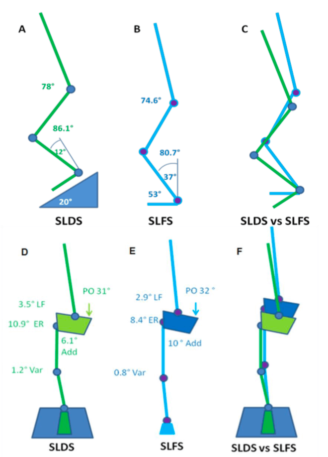
A picture tells a thousand words so in the interests of brevity, the stick squat figure is essentially a summary of two years of work.
So what this means, when someone performs a SLS on a flat surface, relative to a decline surface they tend to have:
- A more upright torso
- More rotation of the pelvis toward the weigh bearing (WB) limb
- Reduced flexion but more adduction and internal rotation of the thigh on the WB hip (pelvic close to femur)
- Less flexion of the knee but the same position relative to the foot as you look from the front (known as frontal plane knee excursion) at the bottom of range
- Reduced internal rotation of the shin
- Reduced ankle flexion
Essentially, in a flat squat you tend to ‘corkscrew’ your pelvis and adopt the medial collapse position much more easily than in the decline squat position. This may because of ankle range of motion issues as well as the ability to adequately recruit pelvic musculature. Yep – two years to get that!
REVISITING THE DATA (2026 RE-ANALYSIS):
Since I first wrote this piece, I have gone back to the original 3D motion capture dataset from my Masters and re-analysed it using more rigorous statistics than I had access to at the time. This work is unpublished and currently being prepared for peer review, so treat it as “watch this space” rather than settled science, but it is a meaningful sharpening of the original findings and, in one respect, a genuine correction to what I wrote above.
The original analysis compared single joint angles at one point in the squat. The re-analysis instead used a multivariate test (Hotelling’s T²) across five hip and pelvis variables together, a false-discovery-rate corrected sweep across eleven segments of the entire kinetic chain (ankle to thorax), and a continuous waveform comparison across the full 0-100% squat cycle rather than a single snapshot.
Two things held up, and one thing did not.
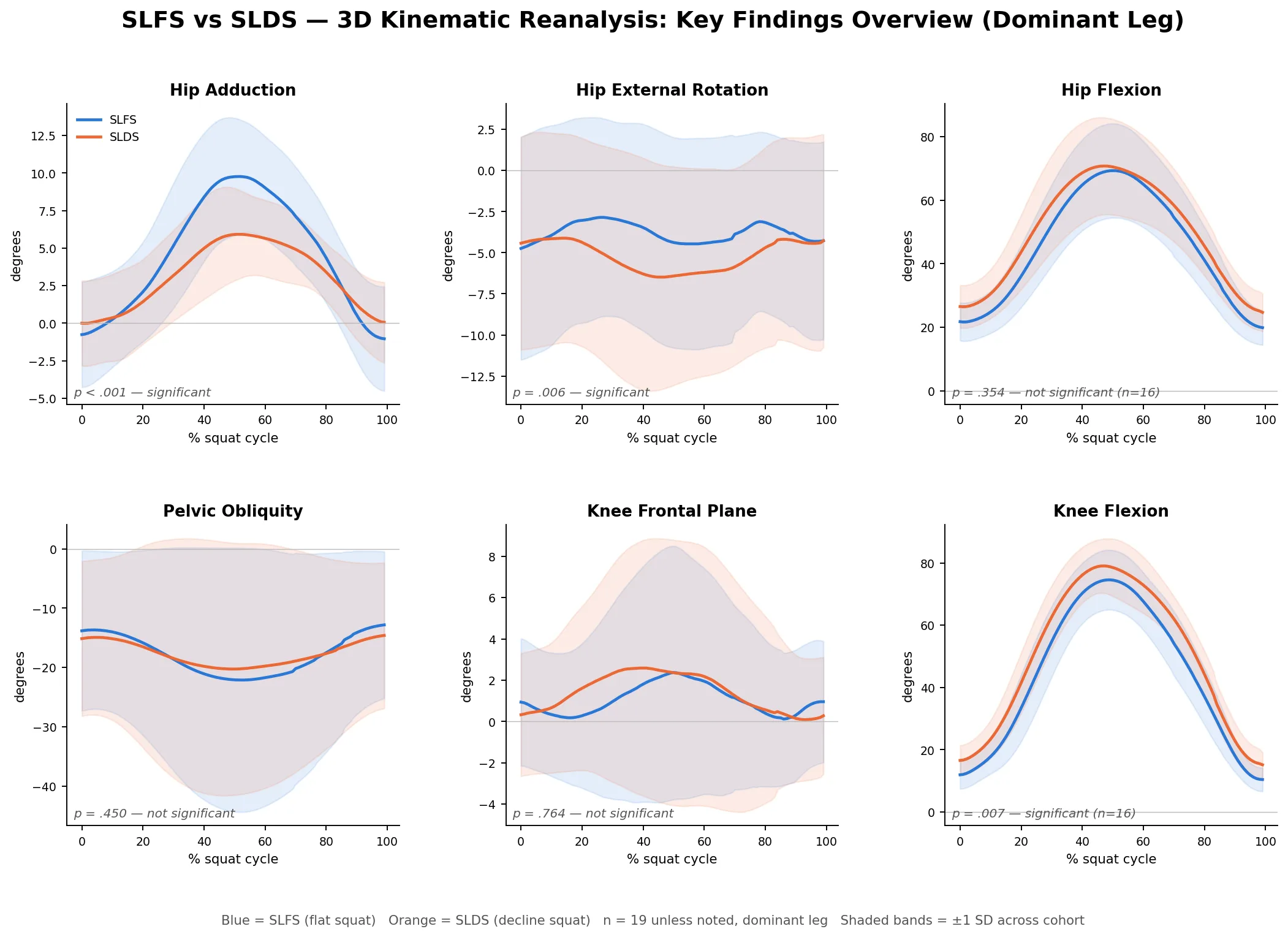
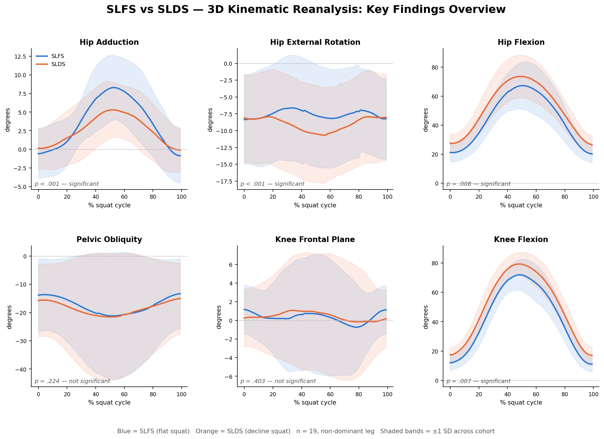
What held up: hip adduction and hip external rotation were consistently greater on the flat squat than the decline squat, by roughly 2-4 degrees on average, and this was robust to the multivariate correction, not just a single-variable finding. Of the eleven segments tested across the whole kinetic chain, only the weight-bearing ankle and the weight-bearing hip reliably differed between conditions (both p<0.001 after FDR correction) - the pelvis, lumbar spine and thorax did not differ. The continuous waveform data also showed that this divergence is not constant through the squat - it emerges gradually through descent and peaks near the deepest point, then closes again on the way up, which matters if you are assessing someone visually and only glance at the very bottom of the movement.
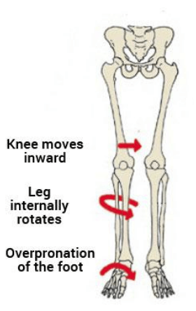
What did not hold up: the “medial collapse” / frontal-plane knee position I described above, comparing flat to decline squat, was not significantly different between the two conditions in this more rigorous analysis. The knee did move differently between the two squat variations, but the difference was in the sagittal (flexion) and transverse (rotation) planes, not the frontal plane. In plain terms, if you are judging flat-versus-decline squat technique by eye and using “does the knee cave in” as your marker, this data suggests that is not actually a variable that distinguishes the two techniques, even though the knee genuinely is moving differently between them in other planes.
Two figures below summarise this: continuous waveform plots (mean ± 1 SD) across the squat cycle for hip adduction, hip external rotation, hip flexion, pelvic obliquity, frontal-plane knee position and knee flexion, for both the dominant and non-dominant limb.