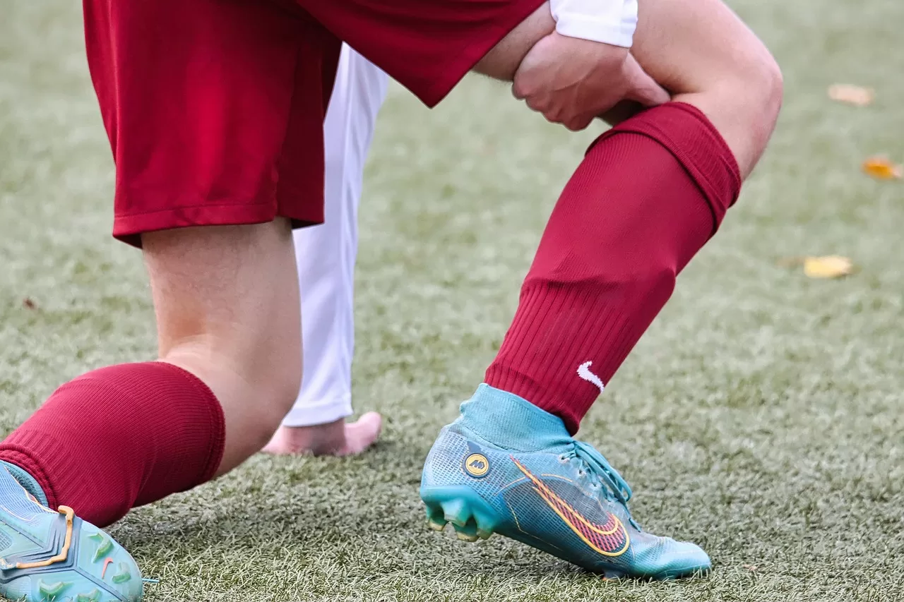
Nordic Curls and the Copenhagen Plank: Two Exercises That Could Save Your Season

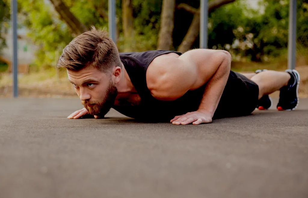
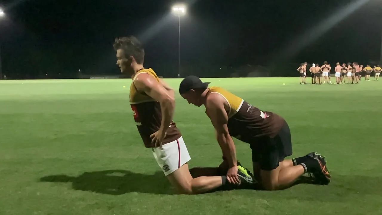
Nordic hamstring curl exercise for hamstring injury prevention

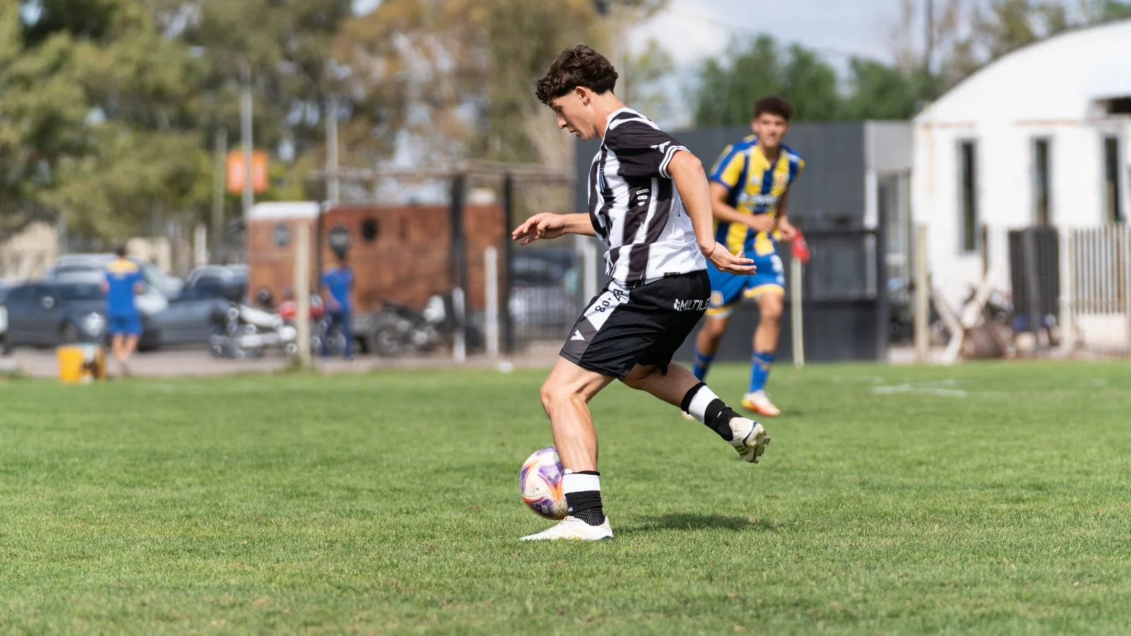
There’s a particular kind of dread that sets in when you feel a twinge sprinting for a ball, or a sharp pull deep in your groin cutting back on the footy field. You know instantly it’s not “just a niggle.” A hamstring or adductor strain has a way of sidelining athletes – weekend warriors and semi-professionals alike – for anywhere from two to twelve weeks, and it has an irritating habit of coming back for round two if the rehab isn’t done properly.

The good news is that a huge amount of research has gone into working out how to stop these injuries before they happen. Two unglamorous, slightly uncomfortable exercises keep coming out on top: the Nordic hamstring curl and the Copenhagen plank. Neither requires fancy equipment. Both have solid evidence behind them. And both are exercises we prescribe regularly here at Praxis for footballers, runners, netballers, and anyone whose sport involves sprinting, kicking, or rapid change of direction.

If you’ve read our piece on hamstring strain injuries and the latest research, you’ll know how frustrating and recurrence-prone these injuries can be. This post is about the flip side – prevention.

WHY THESE TWO EXERCISES?

Hamstring strains and groin/adductor strains are consistently among the most common injuries in field and court sports, and they share a common thread: both muscle groups are asked to produce huge eccentric (lengthening) forces at high speed – the hamstrings decelerating the leg at the end of a sprint stride, the adductors controlling hip abduction during cutting, kicking, and change of direction. Ryan, DeBurca, and Mc Creesh (2014) identified reduced hip adduction strength and previous injury as consistent risk factors for groin and hip injuries in field sports, which is exactly why targeted eccentric strengthening – rather than generic core or gym work – has become the focus of prevention research.

The Nordic hamstring curl trains the hamstrings eccentrically in a lengthened position, closely mimicking the demands of late-swing-phase sprinting. The Copenhagen plank does the same job for the adductors, loading them through a long lever in a side-plank position. Both are simple, both are brutal the first few sessions, and both have research behind them that most gym exercises simply don’t.

THE EVIDENCE FOR NORDIC CURLS

This is where the evidence gets genuinely compelling. Petersen et al. (2011) ran a cluster-randomised controlled trial across 50 Danish soccer teams and found that a progressive Nordic curl programme significantly reduced the rate of both new and recurrent acute hamstring injuries compared with teams doing standard training alone.

That finding has since been confirmed at a much larger scale. Al Attar et al. (2017), in a systematic review and meta-analysis, found that injury prevention programmes including the Nordic hamstring exercise reduced hamstring injury rates by around 51% compared with teams using no such programme. Then van Dyk, Behan, and Whiteley (2019) pooled data from over 8,000 athletes across multiple sports and confirmed an injury rate ratio of roughly 0.49 – essentially, teams doing Nordic curls consistently were about half as likely to suffer a hamstring strain.

There’s also a mechanistic explanation for why it works. Cuthbert et al. (2020) showed that Nordic curl training reliably increases fascicle length in the biceps femoris (the hamstring muscle most commonly injured), meaning the muscle can tolerate greater lengthening at speed before it’s strained – exactly the adaptation you’d want for a sprinting athlete.

WHAT ABOUT THE COPENHAGEN PLANK?

The Copenhagen plank is the adductor equivalent, and the research trail here follows a similar pattern. Ishoi et al. (2016), in a randomised controlled trial with football players, found that an eight-week progressive Copenhagen adduction programme produced large increases in eccentric hip adduction strength – in the order of 9-36% – a strength quality that’s directly linked to groin injury risk.

More importantly, Haroy et al. (2019) tested a Copenhagen-plank-based “Adductor Strengthening Programme” in a cluster-randomised controlled trial across men’s football teams and found it reduced the prevalence of groin problems by roughly 41% over a season. That’s a real-world, injury-outcome level of evidence – not just a strength-testing study.

If you’ve dealt with lingering groin pain before, our article on chronic groin pain and athletic pubalgia covers what happens when adductor-related pain becomes a persistent problem rather than a one-off strain – prevention really is the better option. And for our fast-bowling readers, the same principle of building lengthened-position eccentric capacity applies to the trunk, which is exactly what we discuss in understanding the “side strain” in cricket fast bowlers.

BUSTING THE MYTH: “THESE ARE JUST FOR ELITE FOOTBALLERS”

A common reason these exercises don’t make it into club or weekend-sport training is the assumption they’re only relevant at the professional level. Ironically, Bahr, Thorborg, and Ekstrand (2015) found that even among Champions League and Norwegian Premier League clubs – the exact population the Nordic curl research was built on – only around 11% of teams had fully implemented the protocol, largely due to discomfort, muscle soreness in early weeks, and poor buy-in. If elite clubs with full-time medical staff struggle with compliance, it’s not because the exercise is “too advanced” for amateurs – it’s because dosing and coaching matter, and soreness is manageable with a sensible progression. The upside – roughly halving your hamstring injury risk (van Dyk et al., 2019) – applies to any athlete doing repeated sprinting or change of direction, not just professionals.

HOW TO PROGRAM THEM SAFELY

Both exercises reward patience over bravado. Start with partial-range, assisted Nordic curls (2 sets of 3-5 reps) and low-rep Copenhagen planks (holding from the knee rather than full extension) for the first one to two weeks – expect some genuine delayed-onset soreness early on, which settles quickly with consistent, gradual loading (Cuthbert et al., 2020). From there, progress volume and range over 6-10 weeks toward full-range Nordics and full Copenhagen planks, aiming for two to three sessions a week during pre-season and one maintenance session weekly in-season – mirroring the dosing used in the trials above.

For a visual guide, our physiotherapists have demonstrated the Nordic curl (listed as “Nordic / Hamstring Lower”) on our YouTube channel, @praxisphysiotherapy – well worth a watch before you try it for the first time. The Copenhagen plank is also demonstrated on our YouTube channel, @praxisphysiotherapy.

THE TAKEAWAY

The Nordic hamstring curl and the Copenhagen plank aren’t glamorous, and they’re not comfortable – especially in the first fortnight – but they’re two of the best-evidenced injury prevention exercises in sports medicine. Done consistently and progressed sensibly, they can meaningfully cut your risk of the kind of hamstring or groin strain that ends a season, not just a game.

If you want a strength program tailored to your sport, position, and current niggles rather than a generic YouTube routine, book in with one of our physiotherapists at Praxis. We’ll assess your strength and movement, build a progression that suits your training load, and help you stay on the park.

Until next time, Praxis What You Preach…

📍 Clinics in Teneriffe, Woolloongabba, and Carseldine

💪 Trusted by athletes. Backed by evidence. Here for everyone.

Related Articles
Hamstring Strain Injuries: Lessons from Personal Experience and the Latest Research

Hamstring strains are one of sport’s most common, and most frequently recurring, injuries. Here’s what actually reduces your risk.

Old Man Calf: Why Calf Tears Become So Common After 40

Chasing the football and suddenly felt a pop in your calf? Here’s why this becomes so common as you get older.

Why Your Glutes Aren't Lazy, They're Underdeveloped
Clamshells before squats, banded lateral walks before a run. Here’s why your glute activation routine might be missing the point.
References

Al Attar, W.S.A., Soomro, N., Sinclair, P.J., Pappas, E., & Sanders, R.H. (2017). Effect of Injury Prevention Programs that Include the Nordic Hamstring Exercise on Hamstring Injury Rates in Soccer Players: A Systematic Review and Meta-Analysis. Sports Medicine, 47(5), 907-916.

Bahr, R., Thorborg, K., & Ekstrand, J. (2015). Evidence-based hamstring injury prevention is not adopted by the majority of Champions League or Norwegian Premier League football teams: the Nordic Hamstring survey. British Journal of Sports Medicine, 49(22), 1466-1471.

Cuthbert, M., Ripley, N., McMahon, J.J., Evans, M., Haff, G.G., & Comfort, P. (2020). The Effect of Nordic Hamstring Exercise Intervention Volume on Eccentric Strength and Muscle Architecture Adaptations: A Systematic Review and Meta-analyses. Sports Medicine, 50(1), 83-99.

Haroy, J., Clarsen, B., Wiger, E.G., Oyen, M.G., Serner, A., Thorborg, K., Holmich, P., Andersen, T.E., & Bahr, R. (2019). The Adductor Strengthening Programme prevents groin problems among male football players: a cluster-randomised controlled trial. British Journal of Sports Medicine, 53(3), 145-152.

Ishoi, L., Sorensen, C.N., Kaae, N.M., Jorgensen, L.B., Holmich, P., & Serner, A. (2016). Large eccentric strength increase using the Copenhagen Adduction exercise in football: A randomized controlled trial. Scandinavian Journal of Medicine & Science in Sports, 26(11), 1334-1342.

Petersen, J., Thorborg, K., Nielsen, M.B., Budtz-Jorgensen, E., & Holmich, P. (2011). Preventive Effect of Eccentric Training on Acute Hamstring Injuries in Men’s Soccer: A Cluster-Randomized Controlled Trial. American Journal of Sports Medicine, 39(11), 2296-2303.

Ryan, J., DeBurca, N., & Mc Creesh, K. (2014). Risk factors for groin/hip injuries in field-based sports: a systematic review. British Journal of Sports Medicine, 48(14), 1089-1096.

van Dyk, N., Behan, F.P., & Whiteley, R. (2019). Including the Nordic hamstring exercise in injury prevention programmes halves the rate of hamstring injuries: a systematic review and meta-analysis of 8459 athletes. British Journal of Sports Medicine, 53(21), 1362-1370.

Old Man Calf: Why Calf Tears Become So Common After 40

Runner clutching lower leg in pain after a calf strain“I wasn’t even sprinting…” It’s one of the most common stories I hear in the clinic. “I pushed off to chase the football” “I accelerated after my dog.” “I was only halfway through my run.” “It felt like someone kicked me in the calf.” The diagnosis is usually the same: a calf strain.

In sporting circles, particularly AFL, this injury has become so common that it has earned its own nickname: “Old Man Calf.” The term is tongue in cheek, but the injury certainly isn’t. Calf strains are one of the most frustrating soft-tissue injuries affecting middle-aged athletes and recreational runners. They also have a well documented habit of recurring when they aren’t managed properly.

As someone who treats these injuries regularly, and who has experienced a few myself, I know how debilitating and frustrating they can be. The encouraging part is that they’re also one of the more preventable injuries once you understand why they happen.

Why does it happen?

Age itself isn’t really the problem. What ageing does is create a perfect storm of small physiological changes that gradually chip away at the calf’s ability to tolerate high speed loading.

These changes include a reduction in muscle mass, known as sarcopenia, driven largely by the progressive loss of motor neurons and the muscle fibres they supply (Larsson et al., 2019; Nishikawa et al., 2021). Achilles tendon stiffness also appears to decline with age: research using ultrasound and biofeedback found that older adults produced up to 44% less tendon stiffness than younger adults at matched levels of muscle activation, altering how efficiently force transfers from muscle to bone during push-off (Gray et al., 2024). Interestingly, when load rather than effort is matched, some studies find no real difference in tendon stiffness between younger and older adults, suggesting a good portion of this “stiffness loss” is really a downstream effect of age-related strength loss rather than the tendon tissue itself deteriorating (Jakubowski et al., 2024). Either way, the practical implication is the same: rebuilding strength is central to rebuilding tendon function. Alongside these structural changes, recovery between training sessions slows, maximal strength and power decline, and most people simply stop exposing their calves to genuine sprinting on a regular basis.

None of these changes is dramatic on its own. Together, though, they mean the calf has less “reserve capacity” available when a sudden, explosive movement is demanded of it. That’s why so many people tear a calf doing something they’ve done hundreds of times before without issue.

Why AFL players seem particularly vulnerable

If you’ve watched AFL over the past decade, you’ve probably heard the commentary: “another calf injury,” with veteran players seeming especially susceptible. This isn’t coincidence.

Older AFL players typically retain excellent aerobic fitness and game awareness, but they’re still required to produce repeated maximal accelerations, decelerations and changes of direction, and these explosive actions place enormous force through the calf complex. Data from the AFL’s Soft Tissue Injury Registry back this up. An early analysis of muscle strains across the competition found that age was an independent risk factor for calf strains even after adjusting for injury history, while it wasn’t a risk factor for quadriceps strains, suggesting there’s something specific about how the calf tolerates ageing under repeated high-speed loading (Orchard, 2001). A later 23-year cohort of more than 3,600 muscle strains across the AFL confirmed the pattern, with each additional year of age increasing the odds of a calf strain by around 60% (Orchard et al., 2020). Older players who do sustain a calf strain also tend to take longer to recover (Gray et al., 2025), and a history of calf strain combined with older age substantially raises the risk of a further injury within the following two seasons (Green et al., 2020).

Elite sport doesn’t create this vulnerability; it simply exposes it more publicly.

Recreational runners aren’t immune

You don’t need to play AFL to be at risk. Many of the calf tears seen in clinic occur in park runners, masters athletes, weekend footballers, tennis and pickleball players, golfers walking steep courses, and parents chasing kids around the backyard. Pickleball in particular has seen a sharp rise in soft tissue injuries as participation among older adults has grown, with calf strains featuring among the most commonly recorded injuries at one US orthopaedic centre (Herzberg et al., 2025).

The common thread in all these cases is an athlete who remains active, but who isn’t regularly exposing their calves to high-speed loading. Your cardiovascular fitness might still be excellent. Your calf’s capacity to absorb a sudden sprint or lunge may not be.

Which muscle actually tears?

Detailed anatomical view of the calf musclesMost people simply say they’ve “done their calf,” but several distinct structures can be involved, and it’s worth knowing the difference.

Gastrocnemius is the larger, more superficial of the two major calf muscles. It’s typically injured during explosive acceleration, jumping or sprinting, and pain is often felt high in the calf and occurs suddenly. In a 10-year cohort of elite AFL players, gastrocnemius injuries most commonly affected the medial head (78% of cases), with acceleration being the single most common mechanism of injury (Gray et al., 2025).

Soleus sits deeper and functions more as an endurance muscle. It’s commonly injured during running or prolonged loading, and pain is usually felt lower in the calf, often starting as tightness before becoming more obviously painful. In the same injury registry, soleus injuries were actually more prevalent overall than gastrocnemius injuries, accounting for around 85% of all calf strains in elite AFL players, and were far more likely to recur (Green et al., 2019). Because soleus symptoms tend to develop more gradually, they’re often under-recognised in older runners until the injury is well established.

Why do calf tears keep coming back?

This is probably the biggest source of frustration for patients. Many people feel “pretty good” after three to four weeks: they can walk, they can cycle, and they might even jog comfortably. Unfortunately, healing tissue isn’t the same as fully restored tissue.

The evidence on recurrence is sobering. In elite AFL players, a prior calf strain increased the risk of a further injury several-fold, and older age independently increased that risk further still (Green et al., 2020). A separate analysis of over 3,600 muscle strains across 23 AFL seasons found that the risk of recurrence stays elevated for around 15 weeks after return to play, and that a recent history of the same injury was, by a wide margin, the strongest predictor of injuring it again (Orchard et al., 2020). Put simply, unless strength, endurance, plyometric capacity and sprint exposure are specifically rebuilt, the calf returns to sport with reduced capacity, and eventually something gives again.

Can you prevent “Old Man Calf”?

The encouraging answer is yes. While age can’t be changed, capacity can.

Heavy calf strength. Strong calves tolerate greater force before they fail. Heavy seated and standing calf raises should be part of long-term training, not something reserved for rehab after an injury has already occurred. Eccentric loading in particular has been shown to drive meaningful adaptations in tendon properties and may be especially well suited to older adults, since it can achieve those adaptations at comparatively lower relative demands than other contraction types (Quinlan et al., 2019).

Plyometric training. Skipping, hopping and bounding, performed in small, consistent doses, help the calf improve its ability to store and release elastic energy efficiently.

Runner in starting blocks preparing to sprintSprint exposure. This is perhaps the most overlooked factor of all. Many recreational runners train at a single, moderate pace and then, once every few months, need to sprint for a bus, a ball or a toddler heading for the road. That’s the equivalent of expecting a shoulder to throw a ball at full pace without ever having practised it. Regular controlled strides, accelerations and genuinely faster running help maintain the calf’s resilience to sudden demand.

Consistent training volume. Sharp spikes in training load remain one of the most consistently identified injury risk factors. In professional footballers, a week of unusually high external workload, particularly involving high-intensity accelerations and decelerations, regularly preceded calf strain injuries, and injured players were, on average, older than their uninjured teammates (Soler et al., 2024). The calf tends to prefer gradual, planned increases over heroic weekends bolted onto weeks of inactivity.

Recovery. Sleep, nutrition and ongoing strength training all matter more after 40, simply because recovery isn’t as forgiving as it once was.

Don’t ignore persistent calf tightness

One of the most common mistakes I see is assuming every tight calf just needs stretching. Persistent tightness can actually represent residual weakness, reduced tendon capacity, neural fatigue, or an incompletely healed muscle injury. Stretching alone rarely resolves any of these underlying issues.

A proper assessment should identify why the calf feels tight in the first place. Often, the answer is that it needs to get stronger, not longer.

When should you seek help?

It’s worth having the injury assessed if you experience sudden pain with a popping sensation, difficulty pushing up onto your toes, swelling or bruising, repeated calf strains, or calf tightness that never quite seems to resolve.

A detailed examination can usually determine which structure has been injured, whether imaging is required, and, most importantly, what needs to be rebuilt before you return to running or sport.

The bottom line

“Old Man Calf” might sound like a joke, but it reflects a genuine physiological phenomenon. As we age, our calves gradually lose reserve capacity, and without ongoing strength, power and speed exposure, they’re regularly asked to perform tasks they simply haven’t been prepared for.

Fortunately, this isn’t inevitable. With the right combination of strength training, progressive running, plyometrics and intelligent rehabilitation, many athletes continue running, competing and enjoying sport well into their 50s, 60s and beyond. Age may increase the risk, but it doesn’t have to define your future. If you have struggled (like I have) with calf tears derailing your activity participation, or you fear the return to running stage, book in with one of our physios today to get you back to your best! 

Until next time, Praxis What You Preach 

📍 Clinics in Teneriffe, Buranda, and Carseldine

💪 Trusted by athletes. Backed by evidence. Here for everyone.

Related Articles

Nordic Curls and the Copenhagen Plank: Two Exercises That Could Save Your Season

Two simple exercises, backed by strong evidence, for cutting your hamstring and groin injury risk down significantly.

Shin Splints: Causes, Treatment & How to Get Back to Running Stronger

That nagging ache down your shin isn’t something to just run through. Here’s what’s actually going on, and how to fix it.

Plantar Fasciopathy: Understanding how to heal your heel pain

Heel pain first thing in the morning is one of the most common, and most misunderstood, overuse injuries we see.

References

  1. Larsson L, Degens H, Li M, et al. Sarcopenia: Aging-Related Loss of Muscle Mass and Function. Physiol Rev. 2019;99(1):427-511. DOI
  2. Nishikawa H, Fukunishi S, Asai A, et al. Pathophysiology and mechanisms of primary sarcopenia (Review). Int J Mol Med. 2021;48(2). DOI
  3. Gray AJ, Krupenevich RL, Batsis JA, Sawicki GS, Franz JR. Reduced Achilles tendon stiffness in aging associates with higher metabolic cost of walking. J Appl Physiol. 2024;137(6):1541-1548. DOI
  4. Jakubowski KL, Ludvig D, Lee SSM, Perreault EJ. Aging Does Not Alter Ankle, Muscle, and Tendon Stiffness at Low Loads Relevant to Stance. Ann Biomed Eng. 2024;52(9):2556-2568. DOI
  5. Orchard JW. Intrinsic and extrinsic risk factors for muscle strains in Australian football. Am J Sports Med. 2001;29(3):300-303. DOI
  6. Orchard JW, Chaker Jomaa M, Orchard JJ, et al. Fifteen-week window for recurrent muscle strains in football. Br J Sports Med. 2020;54(18):1103-1107. DOI
  7. Gray C, Pizzari T, Murphy MC, et al. Gastrocnemius Muscle Strain Injury Characteristics in Elite Male Australian Football Players: A 10-Year Longitudinal Cohort Study. J Orthop Sports Phys Ther. 2025;55(10):681-688. DOI
  8. Green B, Lin M, McClelland JA, Semciw AI, Schache AG, Rotstein AH, Cook J, Pizzari T. Return to Play and Recurrence After Calf Muscle Strain Injuries in Elite Australian Football Players. Am J Sports Med. 2020;48(13):3306-3315. DOI
  9. Herzberg SD, Bowman EN, Hill KL. Evaluation of Pickleball-Related Injuries at a Single Institution From 2017 to 2022. Orthop J Sports Med. 2025;13(2). DOI
  10. Green B, Lin M, Schache AG, McClelland JA, Semciw AI, Rotstein A, Cook J, Pizzari T. Calf muscle strain injuries in elite Australian Football players: A descriptive epidemiological evaluation. Scand J Med Sci Sports. 2020;30(1):174-184. DOI
  11. Quinlan JI, Narici MV, Reeves ND, Franchi MV. Tendon Adaptations to Eccentric Exercise and the Implications for Older Adults. J Funct Morphol Kinesiol. 2019;4(3):60. DOI
  12. Soler A, Agulló F, Hernández-Dávó J, Raya-González J, Del Coso J, González-Ródenas J, Moreno-Pérez V. Influence of the External Workload on Calf Muscle Strain Injuries in Professional Football Players: A Pilot Study. Sports Health. 2024;17(1):175-182. DOI
Why Tennis Elbow Takes So Long to Get Better (And What Actually Works)

Why Tennis Elbow Takes So Long to Get Better (And What Actually Works)

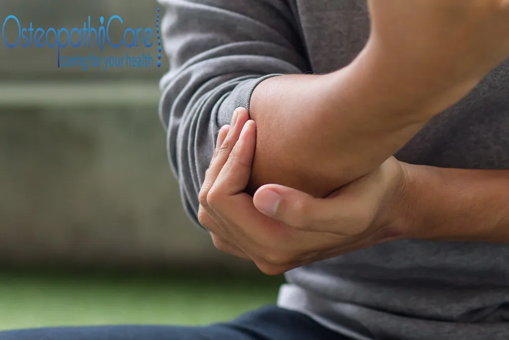
Photo from the Praxis Physiotherapy article: Why Tennis Elbow Takes So Long to Get Better (And What Actually Works)

Ifyou’ve had tennis elbow (also called lateral epicondylalgia), you’ll know it’s not just a “sore elbow.”It’s pain when lifting a coffee cup. Pain shaking hands. Pain turning a door handle. Pain gripping the gym bar or picking up your kids.

And frustratingly, it can hang around for months. So why does tennis elbow take so long to improve? And are injections like cortisone really the quick fix people hope they are?

Let’s unpack it during this installment of Praxis What You Preach

First — It’s Not Really “Inflammation”

Despite the name lateral epicondylitis, most modern research shows that tennis elbow is not primarily an inflammatory condition.

Histological studies consistently demonstrate degenerative tendon changes such as disorganised collagen, increased fibroblasts and vascular changes — rather than classic inflammatory cells. In other words, it’s more accurate to call it a tendinosis or tendinopathy rather than a true “-itis” condition (Herd & Meserve, 2008; Pathan & Sharath, 2023).

This distinction matters. Inflammatory problems (like a sprained ankle in the first few days) often respond quickly to anti-inflammatory treatments.

Degenerative tendon problems do not.

What’s Actually Happening?

Tennis Elbow

In around 90% of cases, the extensor carpi radialis brevis (ECRB) tendon is involved (Pathan & Sharath, 2023). This tendon is heavily loaded during gripping and wrist extension — especially repetitive tasks like typing, manual labour, racquet sports, or gym training.

Over time, repeated micro-loading can exceed the tendon’s capacity. When the tendon fails to adapt effectively, it begins to:

  • Lose collagen organisation
  • Develop microtears
  • Show reduced tensile capacity
  • Become painful with load

This process develops gradually. And that’s one key reason recovery isn’t instant.

“But I’ve Rested It — Why Is It Still Sore?”

Because tendons don’t heal well with complete rest. They need progressive, appropriate load to stimulate remodelling.

Without mechanical stimulus:

  • Collagen alignment worsens
  • Tendon capacity reduces
  • Grip strength declines
  • Pain can actually persist longer

This is why high-quality physiotherapy programs focus on graded strengthening, tendon capacity rebuilding, and load modification, rather than pure rest (Pathan & Sharath, 2023; Yelland et al., 2019).

Natural History: Does It Just Go Away?

Tennis elbow is often described as “self-limiting.” Some reports suggest many cases improve within 8–12 months (Houck et al., 2019), and older literature even suggested high rates of improvement at one year (Herd & Meserve, 2008).

However, that doesn’t mean:

  • It resolves quickly
  • It resolves optimally
  • It resolves without recurrence
  • It resolves without strength deficits

Up to 10% of patients develop persistent symptoms severe enough to consider surgery (Yelland et al., 2019).

And in working adults, particularly manual workers, the functional and economic impact is significant.

So while time helps, structured intervention helps more.

What About Cortisone Injections?

Cortisone injection for tennis elbowCorticosteroid injections are still widely used for tennis elbow. They are:

  • Relatively inexpensive
  • Quick to administer
  • Often very effective for short-term pain relief

And the evidence supports that. Multiple systematic reviews demonstrate that corticosteroid injections are more effective than other treatments in the short term (typically under 12 weeks) (Houck et al., 2019).

The problem? The long term. The same high-level evidence shows that corticosteroid injections:

  • Provide only temporary benefit
  • Are associated with worse long-term outcomes compared to physiotherapy or even a wait-and-see approach (Coombes et al., 2013)
  • Are less effective than platelet-rich plasma (PRP) in the intermediate and longer term (Houck et al., 2019; Kemp et al., 2021)

A 2021 systematic review of systematic reviews concluded:

  • Cortisone = better short-term pain relief
  • PRP = better long-term pain relief and improved function (Kemp et al., 2021)

From a cost-effectiveness perspective, physiotherapy was favoured as a first-line option, while corticosteroid injections showed greater variability and lower probability of being cost-effective over 12 months (Coombes et al., 2013).

Why Might Cortisone Underperform Long Term?

Corticosteroids:

  • Suppress inflammation
  • Reduce pain rapidly
  • Do not improve tendon structure
  • May temporarily weaken tendon tissue

In degenerative tendinopathy, masking pain without improving load capacity can lead to premature return to aggravating activities, and recurrence. For this reason, corticosteroid injections are rarely our first-line strategy at Praxis.

What About PRP?

Platelet-Rich Plasma (PRP) is a biological injection derived from your own blood. It contains concentrated platelets and growth factors intended to stimulate tissue repair.

The evidence is not perfect but it is increasingly supportive in certain contexts.

A large systematic review and meta-analysis found that PRP resulted in improvements exceeding minimal clinically important difference (MCID) thresholds across commonly used outcome measures (VAS, DASH, PRTEE) from 4 weeks through to 104 weeks in many studies (Niemiec et al., 2022).

When compared directly to corticosteroid injections:

  • Cortisone tends to win early
  • PRP tends to win at 3–12 months (Houck et al., 2019; Kemp et al., 2021)

The 2021 review in the International Journal of Sports Physical Therapy concluded that PRP appears to be a more effective long-term treatment option for patients who have failed conservative care (Kemp et al., 2021).

Important Caveats

PRP:

  • Is not a magic bullet
  • Has variability in preparation methods
  • Works best when combined with appropriate load rehabilitation

At Praxis, if PRP is considered, it is typically:

  • For recalcitrant cases
  • After structured rehab has failed
  • Integrated into a progressive strengthening program

So What Actually Works Best?

High-quality evidence consistently supports physiotherapy-directed exercise programs for both short- and long-term improvement (Yelland et al., 2019; Pathan & Sharath, 2023).

Manual therapy combined with exercise has demonstrated meaningful clinical benefits compared to placebo or corticosteroid injection (Yelland et al., 2019).

There is also evidence supporting certain manipulative therapy techniques for symptom reduction (Herd & Meserve, 2008), though exercise remains the cornerstone of recovery.

Importantly, a well-designed physiotherapy program is not just:

  • “Do some wrist curls”
  • “Stretch your forearm”
  • “Wear a brace”

Effective rehab addresses:

  • Tendon load tolerance
  • Grip strength deficits
  • Kinetic chain contributions (shoulder and cervical loading)
  • Work or sport-specific demands
  • Progressive capacity building
  • Assessment of cervical and local nerve contributors to pain and dysfunction

We don’t give away all the details publicly, but it’s far more nuanced than a generic exercise sheet.

Why It Takes Time

Tendons remodel slowly. Unlike muscle tissue, which may respond in weeks, tendon adaptation can take:

  • 8–12 weeks for meaningful structural adaptation
  • 3–6 months for substantial capacity restoration
  • Longer in chronic cases

And if the condition has been present for 6–12 months already, the tissue changes are well established.

Add to that:

  • Poor early management
  • Repeated cortisone injections
  • Ongoing load without strength progression
  • High occupational demands

And recovery timelines extend further.

The Takeaway

Tennis elbow takes time because:

  • It is primarily degenerative, not inflammatory (Herd & Meserve, 2008; Pathan & Sharath, 2023)
  • Tendons adapt slowly
  • Quick fixes often don’t address load capacity
  • Short-term pain relief is not the same as long-term recovery

Cortisone may reduce pain quickly — but does not appear to be the best long-term solution (Houck et al., 2019; Coombes et al., 2013).

PRP shows more promising longer-term outcomes in persistent cases (Niemiec et al., 2022; Kemp et al., 2021) — but still works best when combined with progressive rehabilitation.

Tennis elbow rarely improves by accident — it improves with the right load, at the right time.

If you’re tired of resting, taping, or chasing temporary fixes, our team can guide you through a structured, evidence-based program designed to rebuild tendon capacity and reduce recurrence.

Book an appointment at one of our Brisbane clinics today and start moving forward with a clear plan.

Until next time, Praxis What You Preach…

📍 Clinics in Teneriffe, Buranda, and Carseldine

💪 Trusted by athletes. Backed by evidence. Here for everyone.

Related Articles

Achilles Tendinopathy: How to treat your Achilles Pain

Today on the Praxis What We Preach blog, where we shed light on Achilles tendinopathy, a c…

Rotator Cuff Repair: A Physiotherapy Guide on Recovery and Rehabilitation

Rotator cuff repair is a common surgery for persistent shoulder pain and dysfunction, but …

Jumper’s Knee (Patellar Tendinopathy)

If you play a jumping sport like volleyball, basketball or AFL and feel a nagging pain at …
Rotator Cuff Repair: A Physiotherapy Guide on Recovery and Rehabilitation

Rotator Cuff Repair: A Physiotherapy Guide on Recovery and Rehabilitation

What to Expect from Rotator Cuff Repair: A Physiotherapy Perspective on Recovery and Rehabilitation

Rotator cuff repair surgery is a common and highly effective intervention for individuals suffering from persistent shoulder pain, weakness, or dysfunction due to a torn rotator cuff. Many patients turn to Google with phrases like “rotator cuff surgery recovery timeline,” “shoulder rehab exercises,” or “physiotherapy after shoulder surgery” when looking for answers about what comes next (that may have brought you here!).

While surgical techniques have advanced significantly, the rehabilitation process that follows is equally important in determining a successful outcome. In this Praxis What You Preach blog, we outline what patients can expect from physiotherapy after rotator cuff repair, based on current evidence, clinical best practice and our years of experience dealing with post operative patients.

Phase 1: Protection and Pain Management (Weeks 0–6)

The early stage of rehabilitation focuses on protecting the surgical repair, minimising pain, and reducing inflammation. Patients are typically placed in a shoulder immobiliser or sling for 4–6 weeks to allow early tendon-to-bone healing (Sgroi & Cilenti, 2018; Nikolaidou et al., 2017).

  • Passive Range of Motion (PROM) may begin within this phase under the supervision of a physiotherapist to prevent stiffness while avoiding strain on the healing tendon (Conti et al., 2009).
  • Key goals include:
    • Pain control (using ice, medication, or electrotherapy)
    • Preventing stiffness through gentle PROM in safe planes
    • Maintaining mobility of the elbow, wrist, and hand

“Excessive immobilisation can contribute to shoulder stiffness and muscle atrophy, yet too much movement too soon may compromise tendon healing” (Littlewood et al., 2015).

Phase 2: Controlled Mobilisation (Weeks 7–11)

Once the tendon is more securely integrated with bone, the sling is discontinued and patients begin active-assisted and then active range of motion (AAROM → AROM).

  • Exercises now include:
    • Assisted shoulder flexion and external rotation
    • Scapular control and retraction exercises
    • Isometric strengthening for deltoid and scapular stabilisers

This phase is critical to restoring functional movement without overloading the healing tendon. A slow and structured progression is essential. According to Bandara et al. (2021), protocols that are milestone-based (rather than time-based alone) yield better individualised outcomes.

“Criteria to progress should include pain-free PROM and AROM without compensation or shoulder shrug” (Sgroi & Cilenti, 2018).

Phase 3: Strengthening and Neuromuscular Control (Weeks 12+)

At approximately 12 weeks, patients typically progress to resisted exercises that begin to strengthen the repaired rotator cuff and surrounding musculature. At this stage:

  • Isotonic rotator cuff and scapular muscle training begins
  • Progressive resistance exercises (e.g. banded ER/IR, rows)
  • Incorporation of proprioception and dynamic control (e.g. rhythmic stabilisation, closed-chain activities)

The focus shifts from range of motion to building load tolerance and functional strength. Exercise selection considers tendon healing biology, which shows more mature tendon-to-bone healing around the 12–16 week mark (Nikolaidou et al., 2017; Conti et al., 2009).

“Initiation of functional loading early in the rehabilitation programme does not adversely affect clinical outcome, provided it is gradual and well-monitored” (Littlewood et al., 2015).

Phase 4: Return to Activity and Sport-Specific Rehabilitation (Month 4+)

From four months onwards, many patients begin returning to higher-level tasks depending on their goals:

  • Overhead activities for daily life or sport
  • Plyometric and ballistic loading for athletes
  • Work conditioning or manual labour readiness

At Praxis Physiotherapy, we tailor this phase to your individual goals—whether that’s lifting your toddler, swinging a golf club, or returning to competitive sport.

Some protocols extend formal physiotherapy through months 6–12 for more complex tears or high-functioning individuals.

Communication and Individualisation are Key

Every patient recovers at a different rate depending on:

  • Size and chronicity of the tear
  • Surgical technique
  • Pre-existing stiffness or atrophy
  • Adherence to rehabilitation and exercise

Close collaboration between surgeon, physiotherapist, and patient is essential for long-term success (Sgroi & Cilenti, 2018; Nikolaidou et al., 2017).

“There is strong evidence that early initiation of rehabilitation does not adversely affect clinical outcomes, but should always be individualised” (Littlewood et al., 2015; Bandara et al., 2021).

Final Thoughts 

Rotator cuff repair is only the beginning of the journey. At Praxis Physiotherapy, we provide evidence-based, goal-oriented care from day one post-op through to full return to work, life, and sport.

If you’re preparing for rotator cuff surgery or are currently in recovery, book an appointment at one of our Brisbane locations to begin a structured and personalised rehabilitation program. Begin your recovery the right way.

Until next time, Praxis What You Preach

📍 Clinics in Teneriffe, Buranda, and Carseldine

💪 Trusted by athletes. Backed by evidence. Here for everyone.

Related Articles

Shoulder Stabilisation and Bankart Repair: Your Path Back to Sport

Recovering from a shoulder stabilisation surgery, particularly a Bankart repair, can feel …

ROTATOR CUFF TEARS || Do I Need Surgery?

That age old question in which the answers seems to be becoming increasingly more difficul…

FACT OR FICTION FRIDAY || All rotator cuffs tears need surgery.

Answer: FICTION. The devil is in the detail! The rotator cuff is a group of 4 muscles that…

References

  1. Sgroi, T. A., & Cilenti, M. (2018). Rotator cuff repair: Post-operative rehabilitation concepts. Current Reviews in Musculoskeletal Medicine, 11(1), 86–91. https://doi.org/10.1007/s12178-018-9462-7
  2. Nikolaidou, O., Migkou, S., & Karampalis, C. (2017). Rehabilitation after rotator cuff repair. Open Orthopaedics Journal, 11, 154–162. https://doi.org/10.2174/1874325001711010154
  3. Conti, M., Garofalo, R., & Delle Rose, G. (2009). Post-operative rehabilitation after surgical repair of the rotator cuff. Chirurgia degli Organi di Movimento, 93(Suppl 1), S55–S63. https://doi.org/10.1007/s12306-009-0003-9
  4. Littlewood, C., Bateman, M., Clark, D., Selfe, J., Watkinson, D., Walton, M., & Funk, L. (2015). Rehabilitation following rotator cuff repair: A systematic review. Shoulder & Elbow, 7(2), 115–124. https://doi.org/10.1177/1758573214567702
  5. Bandara, U., An, V. V. G., Imani, S., Nandapalan, H., & Sivakumar, B. S. (2021). Rehabilitation protocols following rotator cuff repair: A meta-analysis of current evidence. ANZ Journal of Surgery, 91(12), 2773–2779. https://doi.org/10.1111/ans.17213

Your Guide to Total Knee Replacement Surgery

Your Guide to Total Knee Replacement Surgery

 Total knee replacement (TKR) is a life-changing procedure for individuals suffering from chronic knee pain, typically caused by osteoarthritis. At Praxis Physiotherapy, we understand that total knee replacement (TKR) surgery is a major decision. As such, we are committed to helping patients navigate their surgical journey and maximize outcomes through tailored prehabilitation and rehabilitation programs.

This guide is designed to walk you through what to expect before and after surgery, how physiotherapy plays a crucial role, and the advanced, evidence-based services we offer to support your journey.

Why Physiotherapy Matters

Physiotherapy isn’t just something you do after surgery—it’s a vital part of your preparation. Prehabilitation (prehab) that begins around six weeks before surgery can improve muscle strength, mobility, and balance, leading to quicker, more successful recoveries post-surgery (Domínguez-Navarro et al., 2020).

Similarly, post-operative physiotherapy supports improved pain relief, better joint function, and faster return to daily activities (Artz et al., 2015), (Fatoye et al., 2021).

Pre-Surgery: Building a Strong Foundation

It’s easy to think, “Why do physio now when the knee is being replaced anyway?” But strengthening and conditioning your body beforehand significantly boosts your post-surgery recovery, helping you get back on your feet faster and with greater confidence. We can address any questions or concerns you may have leading up to the surgery.

Timeline: Ideally begins 6-8 weeks prior to surgery.

Goals:

  • Strengthen muscles around the knee
  • Improve joint mobility
  • Enhance balance and proprioception
  • Educate on post-operative exercises

Key Interventions at Praxis:

  • Reformer Pilates: Our modified prehab programs integrate Pilates to build core stability and lower limb strength. It’s a safe, adaptable way to enhance neuromuscular control before surgery (Levine et al., 2009).
  • Balance Training: Proven to improve post-surgical function when combined with strength training [(Domínguez-Navarro et al., 2020)].
  • Education: We prepare you with strategies to navigate the early post-op period, including mobility aids and pain management.
  • Expert Manual Therapy: Enhances joint mobility, reduces pre-surgical stiffness, and prepares surrounding tissues for optimal post-surgery performance.

Early Post-Op Phase (0-6 weeks)

Immediately following surgery, your primary goals will be managing pain, reducing swelling, and restoring basic mobility.

Many assume recovery only begins once the surgical pain fades—but getting moving early is critical. Guided physiotherapy helps you regain mobility safely, reduce complications, and build confidence from the very start.

Expect:

  • Supervised sessions with focus on safe movement and circulation
  • Gentle range-of-motion and isometric exercises
  • Gait retraining using assistive devices

Evidence-based benefit: Early mobilisation and physiotherapy within days of surgery improve short-term outcomes (Isaac et al., 2005).

Mid to Late Post-Op Phase (6 weeks – 6 months)

At this stage, the intensity of therapy increases to target long-term function. Don’t settle for “good enough” recovery. This phase is where you rebuild your strength, stability, and full mobility—setting the stage for lasting function and confidence in your new joint.

Our Therapeutic Arsenal Includes:

  • Blood Flow Restriction (BFR) Training: Using pneumatic cuffs, we simulate high-load training effects using light resistance. Safe and effective for improving strength post-TKR (Piva et al., 2019).
  • Functional Strength & Balance Training: Tailored to your activity goals.
  • Reformer Pilates: Reactivated in this phase to support low-impact, whole-body conditioning.
  • Access to On-Site Gym Facilities: Ensures continuity and transition from rehab to independent exercise.

Patients receiving a combination of manual therapy and exercise had better functional outcomes than those receiving exercise alone (Karaborklu Argut et al., 2021), a practice we fully embrace at Praxis.

Clinical Expertise You Can Trust

Praxis Physiotherapy works in close collaboration with orthopaedic knee surgeon Dr. Kelly Macgroarty, ensuring a seamless continuum of care. However, we welcome referrals from any orthopaedic surgeon.

You’re not alone in this process. Our experienced team is with you every step of the way—offering expert care, tailored planning, and hands-on support backed by evidence and close collaboration with your surgical team

Our clinicians are highly skilled in post-TKR rehabilitation and stay up-to-date with the latest evidence-based interventions.

What Does the Research Say?

Recent studies underscore the critical value of physiotherapy before and after knee replacement surgery. Prehabilitation, including strength and balance training, has been shown to improve early recovery outcomes [(Domínguez-Navarro et al., 2020)]. Combining manual therapy with exercise yields superior functional gains compared to exercise alone [(Karaborklu Argut et al., 2021), (Abbott et al., 2013)]. Blood Flow Restriction (BFR) training and Pilates have emerged as safe, effective adjuncts to conventional rehabilitation protocols [(Levine et al., 2009), (Piva et al., 2019)]. While short-term improvements in pain and mobility are well-documented, the long-term benefits of physiotherapy interventions vary across studies, highlighting the importance of personalized care and follow-up [(Artz et al., 2015), (Fatoye et al., 2021)].

What Makes Praxis Different?

  • Prehab programs starting 6+ weeks before surgery
  • Use of advanced modalities: BFR cuffs, Reformer Pilates
  • Access to gyms within our clinics
  • Close collaboration with top orthopaedic surgeons
  • One-on-one care tailored to your surgical timeline and goals

Ready to Begin Your Journey?

Total knee replacement doesn’t have to mean months of struggle and guesswork. With the right physiotherapy strategy—starting before your surgery—you can dramatically improve your mobility, reduce pain, and return faster to the activities you love. Reach out to Praxis Physiotherapy today to schedule your pre-operative assessment or post-surgical consultation. Let us guide your recovery with confidence, care, and clinical expertise.

Until next time, Praxis What You Preach…

📍 Clinics in Teneriffe, Buranda, and Carseldine

💪 Trusted by athletes. Backed by evidence. Here for everyone.

References

  • Artz, N. et al. (2015). Effectiveness of physiotherapy exercise following total knee replacement: systematic review and meta-analysis. BMC Musculoskeletal Disorders.
  • Domínguez-Navarro, F. et al. (2020). Preoperative strengthening and balance training. Knee Surgery, Sports Traumatology, Arthroscopy.
  • Fatoye, F. et al. (2021). Clinical and cost-effectiveness of physiotherapy interventions. Archives of Orthopaedic and Trauma Surgery.
  • Karaborklu Argut, S. et al. (2021). Exercise and manual therapy vs exercise alone. PM&R.
  • Levine, B. et al. (2009). Pilates for rehabilitation after total joint arthroplasty. Clinical Orthopaedics and Related Research.
  • Piva, S. et al. (2019). Later-stage exercise vs usual care. JAMA Network Open.
    Praxis Physiotherapy logo

    Visit Us

    CARSELDINE CLUB COOPS
    751 Beams Road
    Carseldine QLD 4034

    TENERIFFE
    91 Commercial Road
    Teneriffe QLD 4005

    WOOLLOONGABBA
    Shop LE201, Level 2
    Centro Buranda Shopping Centre
    Cnr Ipswich Rd & Cornwall Street
    Woolloongabba QLD 4120

    Copyright 2025 Praxis Physiotherapy. All Rights Reserved.

    Related Articles

    Navigating Knee Osteoarthritis: A Physio-Centric Pathway to Strength and Mobility Before Surgery

    Knee osteoarthritis (OA) is one of the most common causes of chronic pain and mobility res…

    Knee Osteoarthritis: Is ‘Bone on Bone’ a painful life sentence?

    Knee Osteoarthritis is a common ailment responsible for pain, loss of function and reduced…

    Capping kneecap pain – Your guide to Anterior Knee Pain (Patellofemoral Pain Syndrome)

    Patellofemoral Pain Syndrome (PFPS) is a common condition that affects the knee joint, par…

    ACL Reconstruction Rehab – Week-by-Week Recovery Guide with Praxis Physio

    ACL Reconstruction Rehab – Week-by-Week Recovery Guide with Praxis Physio

    Overview

    ACL reconstruction surgery marks the beginning of a structured rehabilitation journey. At Praxis Physiotherapy, located in Teneriffe, Carseldine, and Buranda, we provide an evidence-based approach to guide patients from surgery to sport. Backed by over a decade of experience with football teams across Brisbane and collaborative ties with local knee surgeons, our programs are scientifically informed and results-driven.

    Research has shown that a phased, criterion-based rehab plan reduces complication rates and improves return-to-sport outcomes (Shelbourne & Nitz, 1990). The following week-by-week overview reflects current best practice from leading ACL rehab literature.

    Week-by-Week ACL Rehab Milestones

    Prehab: Starting Strong Before Surgery

    If you’re waiting for ACL surgery and your knee has no complicating factors like meniscal locking, there’s good evidence that doing some early rehab — before going under the knife — can significantly improve your recovery trajectory. This phase, often called “prehab,” aims to reduce swelling, restore full knee extension, activate the quadriceps, and build general lower limb strength.

    Research shows that patients who enter surgery with better quadriceps strength and full range of motion recover faster and regain function more effectively post-operatively (Eitzen et al., 2010). In fact, one study in the British Journal of Sports Medicine found that even just 5 sessions of targeted prehab improved early post-op outcomes like walking speed, strength, and self-reported function (Failla et al., 2016).

    Weeks 0–2: Pain, Protection, and Range

    Early rehabilitation begins with swelling and pain management, protection of the graft, and restoration of knee extension. Controlled range-of-motion (ROM) exercises and quadriceps activation, particularly of the vastus medialis, are prioritised. Patients often use crutches to maintain safe gait patterns. Early introduction of blood flow restriction (BFR) training supports muscle maintenance without joint overload (Zazirnyi et al., 2020).

    Checkpoint: Achieve full extension and minimal swelling by Week 2.

    Weeks 2–6: Regain Motion and Begin Strength

    Once inflammation is controlled, attention shifts to regaining full ROM, normalising walking gait, and initiating basic strength exercises such as mini-squats and heel raises. Use of closed kinetic chain exercises is supported for their functional benefit and reduced joint stress (Awad et al., 2017).

    Checkpoint: Full ROM with independent walking and neuromuscular control.

    Weeks 6–12: Strength Foundation

    Patients now begin progressive resistance training using clinic gym equipment, including leg presses, Romanian deadlifts, and lunges. Core strength and dynamic control are emphasised. Light cardio via cycling or elliptical may be introduced. Pilates reformers are utilised at Praxis for controlled joint loading and core development.

    Checkpoint: Strength symmetry reaching 70% of non-injured leg; competent single-leg stance.

    Weeks 12–20: Power and Plyometric Preparation

    This phase involves development of reactive strength and neuromuscular readiness. Jump landing, eccentric hamstring training, and lateral movement patterns are introduced. Key focus is on building capacity for eventual cutting and pivoting movements.

    Checkpoint: Successful hop tests, 80% limb symmetry, and controlled change-of-direction drills.

    Weeks 20–36: Agility and Functional Sport Movements

    Higher-level drills simulate sport-specific movements. Patients perform acceleration/deceleration tasks, direction changes, and reactive decision-making. Plyometrics are progressed in intensity and volume. According to Damian & Damian (2018), phase-specific drills improve psychological readiness and functional return to play (Damian & Damian, 2018).

    Checkpoint: Limb symmetry >90% in strength and hop metrics.

    Months 9–12: Return-to-Sport Preparation

    This stage addresses psychological readiness and simulates sport-specific loading. Functional and fatigue testing are conducted, often including contact drills. Clearance depends on achieving objective strength and control measures (Shelbourne & Patel, 1996).

    Checkpoint: Refer for return-to-sport testing (detailed in a separate blog).

    Practical Insights for Patients

    ACL rehab can be a long and often isolating journey. Many patients report psychological challenges, especially during the early and middle stages when progress may feel slow. At Praxis, we recognise that motivation is a vital part of recovery. Our goal isn’t just to return you to pre-injury levels — it’s to help you become a more resilient, stronger athlete than before. Many ACL injuries stem from non-contact mechanisms linked to strength, control, and movement quality. By targeting these factors throughout rehab, we aim to reduce reinjury risk and elevate athletic performance in the long term.

    ACL rehabilitation is about restoring whole-body function, not just healing a ligament. At Praxis Physiotherapy, we use a combination of BFR, Pilates reformers, and in-house rehab gyms to deliver tailored and progressive programs. Our partnerships with Brisbane’s knee specialists ensure timely updates and coordinated care transitions.

    Why Brisbane Athletes Choose Praxis Physiotherapy

    • Proven protocols developed with elite Brisbane football teams
    • Direct lines of communication with top knee surgeons
    • BFR and Pilates integrated into post-op care
    • Ongoing strength and functional assessments across all phases

    Conclusion

    A structured, evidence-backed approach to ACL rehab dramatically improves recovery outcomes. At Praxis Physiotherapy, we offer a seamless, week-by-week program from day one post-op through to full sport readiness. Our comprehensive model ensures that Brisbane athletes receive the highest standard of care at every stage. If you’re ready to get your rehab underway, book with us! 

    Until next time – Praxis What You Preach

    📍 Clinics in Teneriffe, Buranda, and Carseldine

    💪 Trusted by athletes. Backed by evidence. Here for everyone.


    References

    1. Shelbourne, K.D., & Nitz, P. (1990). Accelerated rehabilitation after anterior cruciate ligament reconstruction. Am J Sports Med.
    2. Zazirnyi, I.M., et al. (2020). Our Point of View at Rehabilitation After ACL Reconstruction. Feofaniya Hospital.
    3. Damian, C. & Damian, M. (2018). Futsal Player Rehabilitation after ACL Reconstruction. Revista Românească.
    4. Awad, O.B. et al. (2017). A Systematic Review of ACL Reconstruction Rehabilitation. Egyptian Journal of Hospital Medicine.
    5. Shelbourne, K.D., & Patel, D.V. (1996). Rehabilitation after autogenous bone-patellar tendon-bone ACL reconstruction. JBJS Am.

    Related Articles

    ACL Rehabilitation: The Role of Physiotherapy in Returning to Life, Activity, and Sport

    Recovering from ACL surgery? Praxis Physiotherapy delivers structured, evidence-based ACL …

    Navigating Knee Osteoarthritis: A Physio-Centric Pathway to Strength and Mobility Before Surgery

    Knee osteoarthritis (OA) is one of the most common causes of chronic pain and mobility res…

    Your Guide to Total Knee Replacement Surgery

    Discover how starting physiotherapy up to six weeks before your knee replacement surgery c…

    From Pitch to Pressing: Leading the Way in Shoulder Pain Recovery

    From Pitch to Pressing: Leading the Way in Shoulder Pain Recovery

    Understanding Rotator Cuff Pain in Active Populations

    Shoulder pain, particularly rotator cuff (RC) injuries, is one of the most common complaints among athletes and active individuals. The rotator cuff, comprising the supraspinatus, infraspinatus, teres minor, and subscapularis, stabilises the shoulder and enables dynamic overhead movement. Athletes in cricket, tennis, AFL, and CrossFit are especially prone to strain this system through repetitive, high-load movements (Desmeules et al., 2025).

    At Praxis Physiotherapy, we specialise in managing rotator cuff injuries with precision. Our expertise spans throwing athletes, tennis players at our Coops Club location, contact sports like AFL, and recreational CrossFit athletes — ensuring tailored care across sporting domains.

    Tailored Treatment Backed by Clinical Evidence

    Current guidelines recommend avoiding over-reliance on imaging and emphasize active rehabilitation. Many rotator cuff cases can be effectively managed without surgery, using structured, progressive rehab programs including resistance training, neuromuscular re-education, and load management (Desmeules et al., 2025).

    Key strategies include:

    • Individualised education about the condition and recovery timeline
    • Isometric and isotonic strengthening of shoulder stabilizers
    • Use of validated outcome tools (e.g., handheld dynamometry, ROM apps) to track progress
    • Selective adjuncts, such as manual therapy or taping, when needed for short-term symptom relief

    In persistent or complex cases, our close collaboration with shoulder and knee surgeon Dr. Kelly Macgroarty ensures a seamless escalation pathway and expert review.

    man throwing yellow, blue, and red Mikasa ballThe Athletic Shoulder: Why Sport-Specific Rehab Matters

    The demands placed on a shoulder in throwing or overhead sports are extreme. During a cricket bowl or tennis serve, angular velocities can reach 7000–7500°/s, and forces on the shoulder joint can exceed bodyweight (Wilk et al., 2009). These actions demand both mobility and stability—a balance referred to as the “thrower’s paradox.”

    Our assessments go beyond the shoulder joint, considering the entire kinetic chain — from trunk control to hip mobility — to ensure optimal movement integration and minimize overload (Cools et al., 2021).

    The Evidence on Rehabilitation & Prognosis

    Recent guidelines emphasize exercise-based rehab as the most effective first-line intervention. Strength gains and symptom reduction are typically seen within 12 weeks if appropriately dosed (Desmeules et al., 2025). Furthermore, the longer pain persists, the lower the likelihood of full recovery from physiotherapy alone (Chester et al., 2013).

    In terms of injury prevention, shoulder-focused warm-up programs — such as FIFA 11+, the Oslo Shoulder Program, and sport-specific throwing drills — have shown a moderate to large effect size in reducing injury risk (Liaghat et al., 2023).

    Return to Sport: Measured, Not Rushed

    Our return-to-sport protocols are designed to ensure both readiness and resilience. We use objective criteria:

    • Strength benchmarks (e.g. ER/IR ratio)
    • Symmetry comparisons
    • Fatigue tolerance testing
    • Sport-specific drills and reactive control

    Whether you’re pressing overhead in a CrossFit WOD, tackling in AFL, or ramping up bowling loads in cricket, our protocols ensure a safe and confident return.

    Prevention: Not an Afterthought

    Our clinic philosophy incorporates injury prevention from the first session (PREVENT | PREPARE | PERFORM). For athletes at our Coops tennis facility, we screen for scapular dyskinesis and GIRD (glenohumeral internal rotation deficit). For CrossFitters, we optimise loading strategies and lifting technique.

    Prevention is an ongoing cycle: screen, intervene, reassess. It’s not just about avoiding injury—it’s about building capacity and sustaining high performance (Cools et al., 2021).

    Conclusion

    Rotator cuff pain doesn’t have to be a long-term setback. With expert diagnosis, individualised rehab, and a sport-specific return plan, most athletes recover without surgery. At Praxis, we combine cutting-edge evidence with clinical experience — and our collaboration with orthopaedic surgeon’s and sports medicine practitioners means you’re in expert hands every step of the way. For more on throwing specifically, check out our blog where we put the Praxis team to the test!

    Ready to take control of your shoulder pain? Book an appointment today and let our team guide you back to strength, confidence, and performance.

    📍 Clinics in Teneriffe, Buranda, and Carseldine

    💪 Trusted by athletes. Backed by evidence. Here for everyone.

    References

    1. Desmeules, F. et al. (2025). Rotator Cuff Tendinopathy: Diagnosis, Nonsurgical Medical Care, and Rehabilitation: A Clinical Practice Guideline. Journal of Orthopaedic & Sports Physical Therapy, 55(4), 235–274.
    2. Wilk, K.E. et al. (2009). Shoulder Injuries in the Overhead Athlete. Journal of Orthopaedic & Sports Physical Therapy, 39(2), 38–54.
    3. Cools, A.M. et al. (2021). The Challenge of the Sporting Shoulder: From Injury Prevention Through Sport-Specific Rehabilitation Toward Return to Play. Annals of Physical and Rehabilitation Medicine, 64, 101384.
    4. Chester, R. et al. (2013). Predicting Response to Physiotherapy for Musculoskeletal Shoulder Pain: A Systematic Review. BMC Musculoskeletal Disorders, 14, 203.
    5. Liaghat, B. et al. (2023). Diagnosis, Prevention, and Treatment of Common Shoulder Injuries in Sport: Grading the Evidence. British Journal of Sports Medicine, 57, 408–416.

    Related Articles

    Rotator Cuff Repair: A Physiotherapy Guide on Recovery and Rehabilitation

    Rotator cuff repair is a common surgery for persistent shoulder pain and dysfunction, but …

    Shoulder Stabilisation and Bankart Repair: Your Path Back to Sport

    Recovering from a shoulder stabilisation surgery, particularly a Bankart repair, can feel …

    Throwing Injuries

    THROWING This week in professional development session, our physio team delved into throwi…

    Strong Bones, Strong Runner: Understanding and Treating Stress Fractures

    Strong Bones, Strong Runner: Understanding and Treating Stress Fractures

    Understanding Stress Fractures in Runners: Risk, Recovery, and Prevention

    Stress fractures are a frustrating reality for many runners. Characterised by small cracks or severe bone reactions due to repetitive load, these injuries can derail training for weeks or months, and in some cases, end seasons or careers. While they are most commonly associated with endurance sports like distance running, the underlying mechanisms are multifactorial and complex. This blog explores the current understanding of stress fractures in runners — including emerging research, rehabilitation strategies, and how to lower your injury risk.

    What Is a Stress Fracture?

    A stress fracture is a type of bone stress injury (BSI), an overuse injury caused by the accumulation of microdamage in bone tissue due to repeated loading. Unlike acute fractures that result from a single traumatic event, stress fractures occur when repetitive sub-threshold forces — like running — outpace the bone’s capacity to repair itself (Hoenig et al., 2022).

    Bone is a dynamic tissue that remodels in response to stress. However, when this remodeling process cannot keep up with microdamage accumulation — due to either an increase in training load or inadequate recovery — bone strength deteriorates. This can progress from a stress reaction to a stress fracture and, if untreated, to a complete fracture (Bergman & Kaiser, 2025; Coslick et al., 2024).

    Medial tibial stress fracture

    Why Are Runners So Prone?

    Running, by nature, imposes repeated high loads on the lower limbs. The tibia (shin bone), metatarsals, femur, and pelvis are frequent stress fracture sites in runners (Hadjispyrou et al., 2023). Several factors contribute to the elevated risk in this group:

    • Training Errors: Rapid increases in volume or intensity, excessive hill work, or high mileage without adequate rest periods.

    • Bone Geometry: Martin & Heiderscheit (2023) found associations between proximal femur geometry and increased stress fracture risk, suggesting that individual anatomical differences can affect how load is distributed through the skeleton.

    • Energy Deficiency: Low energy availability, often associated with disordered eating or high training demands, can impair bone remodeling and increase injury risk — particularly in female athletes.

    • Surface and Footwear: Hard surfaces, old or inappropriate shoes, and poor running biomechanics can all contribute to abnormal load distribution and localized bone stress.

    High-Risk vs Low-Risk Locations

    Not all stress fractures are created equal. According to Coslick et al. (2024), stress fractures are categorized based on location and associated risk of complications:

    • Low-risk sites (e.g., posterior tibia, fibula, second metatarsal shaft) typically heal well with conservative treatment.

    • High-risk sites (e.g., anterior tibia, navicular, femoral neck, and sacrum) are more likely to progress to non-union or full fracture and may require surgical management.

    Photo from the Praxis Physiotherapy article: Strong Bones, Strong Runner: Understanding and Treating Stress Fractures

    A nuanced understanding of the fracture location helps guide both treatment duration and rehabilitation intensity.

    The Cumulative Risk Concept

    Traditional models have viewed stress fractures as the result of isolated risk factors. However, Hamstra-Wright et al. (2021) propose a more integrated concept: the cumulative risk profile. This model acknowledges that risk factors — like energy deficiency, training load spikes, biomechanics, menstrual history, and previous BSIs — rarely occur in isolation.

    In this framework, stress fractures occur when the athlete’s “load capacity” is exceeded by their “training load.” What’s striking is that two runners could follow the same training program but respond very differently based on their individual capacity, bone density, and recovery habits.

    Clinically, this means runners must be assessed holistically. It also underscores the importance of individualized training plans, particularly during return-to-run phases.

    Diagnosis and Imaging

    Early symptoms of a stress fracture include localized pain that worsens with activity and settles with rest. As the injury progresses, pain can persist with walking or even at rest.

    Unfortunately, standard X-rays often miss early bone stress injuries. MRI is the gold standard, able to detect bone marrow edema (early stress reaction) before a fracture line develops (Coslick et al., 2024; Bergman & Kaiser, 2025). Bone scans and CT can also be used in specific cases.

    X ray of periosteal reaction of a stress fracture of the radius

    Rehabilitation and Return to Running

    The cornerstone of stress fracture management is load reduction, typically involving rest from impact activities for 4–8 weeks depending on the site and severity. During this time, runners can usually continue cross-training (e.g., cycling, swimming) to maintain cardiovascular fitness.

    A gradual return-to-run program should be guided by symptom response, starting with walk–run intervals and progressing to continuous running. Strength and conditioning plays a vital role in both rehabilitation and prevention — building muscular resilience to offload bony structures. Calf, hip, and core-focused strength work can significantly reduce recurrence risk and should form part of a comprehensive return-to-run strategy. (You can learn more about how we use strength and conditioning at Praxis Physiotherapy to support our runners here)

    Coslick et al. (2024) emphasises the value of a multidisciplinary approach involving physiotherapists, sports physicians, dietitians, and coaches.

    Running Shoes

    Preventing Stress Fractures: What Runners Can Do

    While not all BSIs are preventable, runners can reduce their risk by addressing modifiable factors:

    • Progress training gradually: Avoid spikes in weekly mileage (>10% per week) and ensure at least one rest day.

    • Fuel adequately: Runners with low energy availability are at significantly increased risk, particularly females with menstrual disturbances.

    • Build strength: Muscle fatigue reduces shock absorption. Strengthening the calves, glutes, and trunk can reduce bone loading.

    • Check your shoes and form: Replace runners every 500–800 km and consider a running gait assessment, especially if you have a history of injury.

    • Listen to your body: Early symptoms like persistent aching, pinpoint bony pain, or pain that lingers after a run shouldn’t be ignored.

    The Bottom Line

    Stress fractures in runners are complex, multifactorial injuries that require a careful balance of training load, nutrition, and recovery. While new imaging and biomechanics research has enhanced our ability to diagnose and understand them, the best approach remains holistic — considering both the runner’s physiology and their environment.

    pair of blue-and-white Adidas running shoes

    At Praxis Physiotherapy, we manage bone stress injuries in athletes of all levels. Whether you’re dealing with your first tibial stress reaction or a sacral stress fracture during marathon prep, we can help guide your recovery and reduce your future risk. Book with us today!

    If you’re interested in how stress fractures affect other athletes — like fast bowlers in cricket — read more our blog on lumbar spine stress fractures here.

    Until next time, Praxis what you Preach

    📍 Clinics in Teneriffe, Buranda, and Carseldine

    💪 Trusted by athletes. Backed by evidence. Here for every body. 

    References

    Bergman, R., & Kaiser, K. (2025). Stress Reaction and Fractures. In StatPearls. StatPearls Publishing. Retrieved from

    Coslick, A. M., Lestersmith, D., Chiang, C. C., Scura, D., Wilckens, J. H., & Emam, M. (2024). Lower extremity bone stress injuries in athletes: An update on current guidelines. Current Physical Medicine and Rehabilitation Reports, 12(1), 39–49.

    Hamstra-Wright, K. L., Huxel Bliven, K. C., & Napier, C. (2021). Training load capacity, cumulative risk, and bone stress injuries: A narrative review of a holistic approach. Frontiers in Sports and Active Living, 3, 665683.

    Hadjispyrou, S., Hadjimichael, A. C., Kaspiris, A., Leptos, P., & Georgoulis, J. D. (2023). Treatment and rehabilitation approaches for stress fractures in long-distance runners: A literature review. Cureus, 15(11), e49397.

    Hoenig, T., Ackerman, K. E., Beck, B. R., Bouxsein, M. L., Burr, D. B., Hollander, K., Popp, K. L., Rolvien, T., Tenforde, A. S., & Warden, S. J. (2022). Bone stress injuries. Nature Reviews Disease Primers, 8, 26.

    Martin, J. A., & Heiderscheit, B. C. (2023). A hierarchical clustering approach for examining the relationship between pelvis–proximal femur geometry and bone stress injury in runners. Journal of Biomechanics, 160, 111782.

    Related Articles

    Stress Fractures in Young Cricket Fast bowlers: A persistent challenge

    Lumbar stress fractures are one of the most serious and persistent injuries affecting cric…

    Shin Splints: Causes, Treatment & How to Get Back to Running Stronger

    Key Takeaways Shin splints (MTSS) are an overuse bone stress injury. Training load errors …

    Why lifting is your missing endurance link: A guide for long distance runners (Part 1)

    You have the shoes, the GPS watch, training schedule and alarm set for 5am. You are dedica…

    Shoulder Stabilisation and Bankart Repair: Your Path Back to Sport

    Shoulder Stabilisation and Bankart Repair: Your Path Back to Sport

    Recovering from a shoulder stabilisation surgery, particularly a Bankart repair, can feel like navigating a winding trail. At Praxis Physiotherapy, we understand that athletes don’t just want to heal – they want to return stronger, more confident, and ready for action. This blog explores the key milestones in overhead and contact sport rehabilitation and the compelling evidence that supports structured physiotherapy.

    Understanding Shoulder Stabilisation and Bankart Repair

    The Bankart repair is a surgical intervention for traumatic anterior shoulder dislocations, which are most common in young, active populations – particularly those involved in contact or overhead sports. This procedure reattaches the torn labrum to the glenoid, restoring joint stability. While the surgery addresses structural instability, it is only the first step. Rehabilitating the shoulder to perform under high-stress, dynamic sporting conditions is where physiotherapy becomes crucial (Coyle et al., 2022).


    Rehabilitation Phases: Beyond the Basics

    Rehabilitation after Bankart repair generally progresses through four overlapping phases:

    1. Protection & Early Mobility (0–6 weeks)

    Initial goals include reducing pain and inflammation while protecting the repair. Gentle passive and assisted range-of-motion exercises begin, with sling use gradually tapered.

    2. Strength Building (6–12 weeks)

    Isometric and light resistance training begins. Scapular control and rotator cuff strengthening are vital. Coyle et al. (2022) found wide variability in when strengthening begins, from 1 to 12 weeks, underscoring the importance of tailored plans.

    3. Advanced Control & Load Tolerance (12–20 weeks)

    This phase introduces overhead activity simulation, plyometrics, and proprioceptive drills. Neuromuscular training improves shoulder resilience, especially under rapid direction changes and contact stress (Ialenti et al., 2017).

    4. Return to Sport (20+ weeks)

    Athletes progressively re-engage in sport-specific drills, initially non-contact, then full-contact scenarios. Full return to competitive play often occurs around 5–6 months, but timelines vary based on sport demands (Kasik et al., 2019).


    The Evidence: Why Physiotherapy Matters

    • A systematic review by Rossi et al. (2021) revealed that 27% of athletes failed to return to sport post-surgery. Most cited fear of reinjury, not physical limitation, as the main barrier. Targeted rehab can address both physical readiness and confidence.
    • Kim et al. (2023) showed that factors such as shoulder strength, proprioception, and psychological readiness were predictive of successful return. Structured physiotherapy addresses all three.
    • The American Journal of Sports Medicine confirms that progressive loading, especially for overhead and contact tasks, enhances long-term outcomes and reduces recurrence rates (Kim et al., 2023).
    • Alsomali et al. (2021) and Stone & Pearsall (2014) agree that sport-specific milestones are crucial, with a general return-to-contact timeline of 16–24 weeks depending on the sport.

    Back in the Game: What Sets Our Approach Apart

    At Praxis Physiotherapy, our shoulder rehabilitation programs don’t just follow protocols – they evolve with the athlete. We incorporate evidence-based practices and tailor each phase to your sport, position, and performance goals. Whether you’re a rugby forward or a volleyball setter, our rehab plan adapts to your demands.

    We emphasise:

    • Early and progressive exposure to overhead mechanics
    • Integrated neuromuscular training for dynamic stability
    • Gradual and safe return-to-contact drills
    • Psychological readiness assessments to overcome fear of reinjury


    Summary Timeline for Return to Sport

    Phase Timeframe Focus
    Protection & Early Mobility 0–6 weeks Pain control, protected motion
    Strength & Motor Control 6–12 weeks Rotator cuff & scapular strengthening
    Overhead & Contact Prep 12–20 weeks Plyometrics, proprioception, advanced drills
    Return to Sport 20–26+ weeks Gradual return to contact and full intensity

    Final Thoughts: Your Comeback Starts with the Right Team

    Recovering from shoulder stabilisation surgery is not just about healing – it’s about coming back better. Evidence clearly shows that structured, progressive physiotherapy is essential for returning to sport safely and confidently.

    At Praxis Physiotherapy, we’re here to guide that journey every step of the way. If you are ready to get started, book online today.

    Until next time, Praxis What You Preach…

    📍 Clinics in Teneriffe, Buranda, and Carseldine

    💪 Trusted by athletes. Backed by evidence. Here for everyone.

    References

    • Coyle, M., Jaggi, A., Weatherburn, L., Daniell, H., & Chester, R. (2022). Post-operative rehabilitation following traumatic anterior shoulder dislocation: A systematic scoping review. Shoulder & Elbow, 15(5), 554–565.
    • Ialenti, M. N., Mulvihill, J. D., Feinstein, M., Zhang, A. L., & Feeley, B. T. (2017). Return to play following shoulder stabilization: A systematic review and meta-analysis. Orthopaedic Journal of Sports Medicine, 5(9)
    • Kasik, C. S., Rosen, M. R., Saper, M. G., & Zondervan, R. L. (2019). High rate of return to sport in adolescent athletes following anterior shoulder stabilisation: A systematic review. Journal of ISAKOS, 4(1), 43–50.
    • Kim, M., Haratian, A., Fathi, A., Kim, D. R., Patel, N., Bolia, I. K., … & Weber, A. E. (2023). Can we identify why athletes fail to return to sports after arthroscopic Bankart repair? A systematic review and meta-analysis. The American Journal of Sports Medicine, 51(9), 2480–2486.
    • Rossi, L. A., Tanoira, I., Brandariz, R., Pasqualini, I., & Ranalletta, M. (2021). Reasons why athletes do not return to sports after arthroscopic Bankart repair: A comparative study of 208 athletes with minimum 2-year follow-up. Orthopaedic Journal of Sports Medicine, 9(7)
    • Alsomali, K., Kholinne, E., Nguyen, T. V., Cho, C.-H., Kwak, J.-M., Koh, K.-H., & Jeon, I.-H. (2021). Outcomes and return to sport and work after open Bankart repair for recurrent shoulder instability: A systematic review. Orthopaedic Journal of Sports Medicine, 9(10)
    • Stone, G. P., & Pearsall, A. W. (2014). Return to play after open Bankart repair: A systematic review. Orthopaedic Journal of Sports Medicine, 2(2),

    Related Articles

    Rotator Cuff Repair: A Physiotherapy Guide on Recovery and Rehabilitation

    Rotator cuff repair is a common surgery for persistent shoulder pain and dysfunction, but …

    From Pitch to Pressing: Leading the Way in Shoulder Pain Recovery

    Struggling with shoulder pain that’s holding you back? Learn how athletes can overcome rot…

    Throwing Injuries

    THROWING This week in professional development session, our physio team delved into throwi…
    Pain in the Neck: Why Your Neck Hurts and What To Do About It

    Pain in the Neck: Why Your Neck Hurts and What To Do About It

    Neck pain is one of the most common reasons people seek physiotherapy – and for good reason. Whether it creeps in during long days at the desk, flares after a tough workout, or simply starts for no apparent reason, it can become an ongoing source of discomfort and limitation.

    At Praxis Physiotherapy, we see patients every week with neck pain ranging from occasional stiffness to chronic, persistent aches. The good news? Physiotherapy – particularly manual therapy and targeted exercise – can make a real difference.

    So, What Causes Neck Pain?

    Most neck pain we treat is classed as “non-specific neck pain” (Verhagen 2021; Almalki et al. 2024). That means it doesn’t come from a single clear source like a fracture or disc bulge, but rather a combination of mechanical, postural, and sometimes psychosocial factors.

    Risk factors include:

    • Prolonged static or awkward postures (like slouching over a desk)
    • High computer use (>75% of the workday)
    • Stress, anxiety, poor sleep or low mood
    • Lack of physical activity or poor muscle endurance (Cagnie et al. 2007; Louw et al. 2017)

    Side view of senior man holding neck with visible discomfort, highlighting neck pain relief.

    Importantly, neck pain often fluctuates – it might settle for weeks or months before flaring again. Up to 70% of people will experience neck pain in their lifetime, and around half of those will go on to experience recurring or chronic symptoms (Osborne et al. 2024).

    What Actually Helps?

    Let’s get straight to it. Here’s what the research says works – and what doesn’t.

    Targeted Strengthening Exercises

    A recent meta-analysis by Louw et al. (2017) showed strengthening exercises are consistently more effective than doing nothing. These exercises improve both pain and quality of life for office workers with non-specific neck pain.

    Chen et al. (2018) reinforced this, finding the biggest improvements came from neck/shoulder-specific strength work done consistently. The same review highlighted that those who stuck to their program got the best results — a helpful reminder that consistency trumps intensity.

    Interestingly, Osborne et al. (2024) found neck-specific resistance training not only helped pain but also changed how the nervous system processed pain – reducing hypersensitivity measured by QST (quantitative sensory testing). That’s not just “feeling better” – it’s a measurable shift in how your body interprets threat and discomfort.

    Manual Therapy (With Exercise)

    Close-up of a therapist giving a relaxing shoulder massage, enhancing wellness and stress relief.

    Cervical and thoracic mobilisations – particularly when paired with exercise – help reduce pain and restore movement (Verhagen 2021; Damgaard et al. 2013). At Praxis, we’ll often use hands-on techniques in the early phase to loosen stiff joints or reduce muscle guard

    ing, before layering in exercise to drive long-term change.

    Manual therapy alone can offer short-term relief, but it’s the combination with exercise that produces meaningful, sustained improvement.

    Close-up of woman using blue massage balls for neck relief against a wall.A Multimodal Approach

    Combining manual therapy, strengthening, posture coaching, and education works better than relying on just one of these (Damgaard et al. 2013). This reflects our whole-person approach at Praxis – treating not just the neck, but the patterns, habits, and loads that contribute to the issue.

    What About Stretching?

    Stretching can feel good – and sometimes helps with short-term symptom relief – but strengthening is where the real long-term benefit lies (Louw et al. 2017). That said, we’ll often include mobility work alongside strengthening in the early phases of your rehab, especially if movement is limited or provoking.

    And What Doesn’t Help?

    Unfortunately, there’s still a lot of outdated advice and overreliance on passive treatments. Prolonged rest, neck braces, or relying solely on massage or dry needling – without addressing strength, posture, or movement – rarely produce lasting results.

    Electrotherapy or ultrasound alone have limited evidence (Damgaard et al. 2013), and while they may provide short-term comfort, they don’t improve long-term function or resilience.

    What You Can Expect at Praxis

    Your physio will:

    1. Take a thorough history and assessment to rule out anything serious.
    2. Identify movement deficits, strength imbalances, or aggravating work setups.
    3. Use manual therapy to settle symptoms and restore range of motion.
    4. Build a personalised exercise plan focused on restoring strength and endurance.
    5. Offer ergonomic and postural coaching to help you load your neck better, not less.

    Whether you’re a desk-bound professional, a busy parent, or an elite athlete – your neck pain deserves proper, evidence-based care.

    Struggling with neck pain that just won’t go away? Let one of our experienced physios at Praxis guide you back to feeling and moving better – book today

    Until next Praxis What You Preach..

    📍 Clinics in Teneriffe, Buranda, and Carseldine
    💪 Trusted by athletes. Backed by evidence. Here for everyone.

    Related Articles

    Why The “Wait And See” Approach May Leave You With A Poorer Outcome.

    We are all guilty of it. Putting off seeing someone about that niggle. “it will be r…

    FACT OR FICTION – I’m in pain, I’m damaging something

    FICTION! Stay with me on this one as it can be confusing. What Is Pain, Really?Pain = An u…

    The Benefits of Remedial Massage

    Remedial Massage — Indulgence or Evidence-Based Care? We all enjoy the occasional indulgen…